The thought of an interview can be nerve-wracking, but the right preparation can make all the difference. Explore this comprehensive guide to Neuroanatomy and Neurophysiology interview questions and gain the confidence you need to showcase your abilities and secure the role.
Questions Asked in Neuroanatomy and Neurophysiology Interview
Q 1. Describe the major divisions of the brain and their functions.
The brain is broadly divided into three major parts: the cerebrum, cerebellum, and brainstem. Each plays a crucial, distinct role in our overall functioning.
- Cerebrum: This is the largest part, responsible for higher-level cognitive functions. It’s divided into two hemispheres (left and right), each controlling the opposite side of the body. Specific regions within the cerebrum, called lobes (frontal, parietal, temporal, and occipital), specialize in different tasks. For instance, the frontal lobe is crucial for planning and decision-making, the parietal lobe processes sensory information, the temporal lobe handles auditory processing and memory, and the occipital lobe interprets visual information.
- Cerebellum: Located beneath the cerebrum, the cerebellum primarily coordinates movement, balance, and posture. It doesn’t initiate movement but fine-tunes it, ensuring smooth, coordinated actions. Think of it as the brain’s ‘autopilot’ for motor control. Damage to the cerebellum can lead to tremors, ataxia (loss of coordination), and difficulties with balance.
- Brainstem: This connects the cerebrum and cerebellum to the spinal cord. It controls essential life-sustaining functions such as breathing, heart rate, and sleep-wake cycles. The brainstem is comprised of the midbrain, pons, and medulla oblongata, each with specific roles in regulating these vital processes. Damage to the brainstem can have life-threatening consequences.
Imagine a conductor leading an orchestra: the cerebrum is the conductor making the strategic decisions; the cerebellum is the section leader ensuring each instrument plays in harmony; and the brainstem is the essential power source keeping the orchestra running.
Q 2. Explain the process of synaptic transmission.
Synaptic transmission is the process by which neurons communicate with each other. It involves the transmission of a signal (neurotransmitter) across a tiny gap called the synapse, from the presynaptic neuron (sender) to the postsynaptic neuron (receiver).
- Action Potential Arrival: An electrical signal, called an action potential, travels down the axon of the presynaptic neuron.
- Neurotransmitter Release: When the action potential reaches the axon terminal, it triggers the release of neurotransmitters stored in vesicles. These vesicles fuse with the presynaptic membrane and release their contents into the synaptic cleft (the gap between neurons).
- Neurotransmitter Binding: The released neurotransmitters diffuse across the synaptic cleft and bind to specific receptor proteins located on the postsynaptic neuron’s dendrites.
- Postsynaptic Potential: This binding triggers a change in the postsynaptic neuron’s membrane potential, which can either be excitatory (depolarizing, making the neuron more likely to fire an action potential) or inhibitory (hyperpolarizing, making the neuron less likely to fire).
- Neurotransmitter Removal: The neurotransmitters are then quickly removed from the synaptic cleft through reuptake by the presynaptic neuron, enzymatic degradation, or diffusion. This ensures that the signal is brief and precisely controlled.
Think of it like a key (neurotransmitter) fitting into a lock (receptor). The turning of the key (binding) triggers an action (postsynaptic potential). The removal of the key ensures the lock can be used again.
Q 3. What are the different types of glial cells and their roles?
Glial cells, often called neuroglia, are non-neuronal cells in the central nervous system (brain and spinal cord) and the peripheral nervous system. They provide structural support, insulation, and nourishment to neurons. There are several types:
- Astrocytes: These star-shaped cells are the most abundant glial cells. They provide structural support, regulate the chemical environment around neurons, form the blood-brain barrier, and contribute to synaptic transmission.
- Oligodendrocytes (CNS) and Schwann cells (PNS): These cells produce myelin, a fatty substance that insulates axons and speeds up nerve impulse conduction. Oligodendrocytes myelinate multiple axons in the CNS, while Schwann cells myelinate a single axon segment in the PNS.
- Microglia: These are the resident immune cells of the CNS. They act as scavengers, removing cellular debris, pathogens, and damaged neurons.
- Ependymal cells: These cells line the ventricles (fluid-filled cavities) of the brain and spinal cord. They produce and circulate cerebrospinal fluid (CSF), which cushions and protects the brain and spinal cord.
Imagine a city: neurons are the buildings, astrocytes are the city’s infrastructure and sanitation, oligodendrocytes/Schwann cells are the electrical wiring, microglia are the police force, and ependymal cells are the water management system.
Q 4. Describe the function of the blood-brain barrier.
The blood-brain barrier (BBB) is a highly selective semipermeable border of endothelial cells that prevents many substances from entering the brain’s extracellular fluid. This protects the delicate brain tissue from harmful substances in the bloodstream while allowing essential nutrients and oxygen to pass through.
The BBB is composed of tightly joined endothelial cells lining the brain’s capillaries, supported by astrocytic end-feet. This tight junction prevents the paracellular passage of many molecules. Specific transport mechanisms, such as carrier proteins and receptor-mediated transcytosis, are responsible for the selective transport of essential molecules into the brain. The BBB is crucial in maintaining a stable brain environment and protecting against toxins, pathogens, and fluctuations in blood composition.
For example, many drugs have difficulty crossing the BBB, which is a significant challenge in developing treatments for neurological disorders. This selectivity is a double-edged sword; while protecting the brain, it also limits drug delivery.
Q 5. Explain the difference between EEG and EMG.
Both EEG and EMG are electrophysiological techniques used to measure electrical activity in the body, but they target different systems:
- Electroencephalography (EEG): Measures the electrical activity of the brain using electrodes placed on the scalp. It detects the summed postsynaptic potentials of cortical neurons and provides information about brain states (e.g., sleep, wakefulness, seizures). EEG signals are relatively low in amplitude and show a variety of wave patterns (alpha, beta, delta, theta) that vary based on brain state and activity.
- Electromyography (EMG): Measures the electrical activity of muscles using electrodes placed on the skin or inserted into the muscle. It detects the action potentials generated by muscle fibers during contraction and provides information about muscle function, nerve conduction, and neuromuscular disorders. EMG signals are of higher amplitude than EEG signals and are directly related to muscle activation.
In essence, EEG looks at the brain’s electrical chatter, while EMG listens to the muscle’s electrical signals. Both are valuable diagnostic tools in neurology.
Q 6. What are the key components of a reflex arc?
A reflex arc is a neural pathway that mediates a reflex action. A reflex is an involuntary and nearly instantaneous movement in response to a stimulus. The key components are:
- Receptor: A sensory receptor detects a stimulus (e.g., touch, heat, pain).
- Sensory Neuron (Afferent Neuron): This neuron transmits the signal from the receptor to the spinal cord or brainstem.
- Integration Center: This is typically a synapse in the spinal cord or brainstem, where the sensory neuron’s signal is processed.
- Motor Neuron (Efferent Neuron): This neuron carries the signal from the integration center to the effector.
- Effector: This is a muscle or gland that responds to the signal (e.g., muscle contraction, gland secretion).
Example: The knee-jerk reflex. Tapping the patellar tendon stretches the muscle, activating muscle spindles (receptors). Sensory neurons transmit the signal to the spinal cord, where it synapses with motor neurons. Motor neurons stimulate the muscle to contract, causing the knee to jerk.
Q 7. Describe the pathway of visual information processing.
Visual information processing begins with the eyes and continues through a complex series of neural pathways in the brain. The process can be summarized as follows:
- Retina: Photoreceptor cells (rods and cones) in the retina convert light into electrical signals.
- Optic Nerve: These signals are transmitted via the optic nerve to the optic chiasm.
- Optic Chiasm: Here, fibers from the nasal (inner) half of each retina cross over to the opposite side of the brain. This ensures that information from both eyes is processed together in the same hemisphere.
- Optic Tract: The fibers continue as the optic tract to the lateral geniculate nucleus (LGN) of the thalamus.
- Lateral Geniculate Nucleus (LGN): This acts as a relay station, processing and relaying visual information to the primary visual cortex.
- Primary Visual Cortex (V1): Located in the occipital lobe, this area is responsible for initial visual processing, such as edge detection and orientation sensitivity.
- Extrastriate Cortex: Visual information is then processed in different areas of the extrastriate cortex (V2, V3, V4, V5 etc.), specialized for different aspects of vision, such as color, motion, and form.
- Higher Visual Areas: Finally, visual information reaches higher visual areas involved in object recognition, spatial awareness, and other complex visual functions.
Imagine a pipeline carrying information: The retina collects the raw data, the optic nerve transmits it, the LGN sorts it, the visual cortex processes it, and higher areas make sense of it all. Each step is crucial for a complete and coherent visual experience.
Q 8. Explain the role of the cerebellum in motor control.
The cerebellum, often called the “little brain,” plays a crucial role in coordinating voluntary movement, maintaining balance, and regulating muscle tone. It doesn’t initiate movement; rather, it refines and coordinates movements initiated by the motor cortex. Think of it like a highly skilled editor, ensuring the movements are smooth, precise, and accurate.
The cerebellum receives input from various sources, including the motor cortex (informing it of intended movements), sensory receptors (providing feedback on actual movement), and the vestibular system (monitoring balance). It processes this information and sends corrective signals back to the motor cortex, helping to adjust ongoing movements and ensure accuracy. For example, imagine trying to throw a dart. The motor cortex initiates the throwing motion, but the cerebellum fine-tunes the trajectory, ensuring the dart hits the target. Damage to the cerebellum can lead to ataxia, characterized by clumsy, uncoordinated movements, tremors, and difficulties with balance.
Q 9. What are the different types of sensory receptors and their modalities?
Sensory receptors are specialized cells that detect various stimuli and convert them into electrical signals that the nervous system can understand. Different types of receptors respond to different modalities (types of stimuli).
- Mechanoreceptors: Respond to mechanical pressure or distortion. Examples include those in the skin (detecting touch, pressure, vibration), muscles (muscle spindles sensing muscle length), and inner ear (detecting sound and balance).
- Thermoreceptors: Detect temperature changes. Separate receptors exist for hot and cold.
- Nociceptors: Respond to noxious stimuli that cause pain. These can detect chemical, mechanical, or thermal damage.
- Chemoreceptors: Respond to chemical stimuli. These include taste buds (detecting sweet, sour, salty, bitter, umami), olfactory receptors in the nose (detecting smells), and receptors in blood vessels (monitoring oxygen and carbon dioxide levels).
- Photoreceptors: Respond to light. Rods and cones in the retina are examples, responsible for vision.
Each type of receptor has a specific receptive field, the area of the body or environment it monitors. The intensity and location of the stimulus are encoded in the frequency and pattern of action potentials generated by the receptors.
Q 10. Describe the neurophysiological basis of pain.
The neurophysiological basis of pain is complex, involving multiple pathways and brain regions. It’s not simply a direct response to tissue damage but rather a multifaceted sensory and emotional experience.
Nociceptors, the pain receptors, are activated by noxious stimuli. They transmit signals via peripheral nerves to the spinal cord. These signals then travel up the spinal cord via the spinothalamic tract to the brainstem, thalamus, and eventually various areas of the cortex, including the somatosensory cortex (processing sensory aspects of pain), the anterior cingulate cortex (processing the emotional aspects of pain), and the prefrontal cortex (involved in cognitive evaluation of the pain experience).
The experience of pain is modulated by descending pathways from the brain that can inhibit or amplify pain signals. This is why factors like stress, emotions, and expectations can influence how we perceive pain. Endorphins and other endogenous opioids play a significant role in pain modulation. For example, during strenuous exercise, the release of endorphins can alleviate pain.
Q 11. Explain the process of long-term potentiation (LTP).
Long-term potentiation (LTP) is a persistent strengthening of synapses based on recent patterns of activity. It’s a cellular mechanism underlying learning and memory. Essentially, it’s a strengthening of the connection between two neurons, making it easier for them to communicate with each other in the future.
LTP typically occurs at excitatory synapses that use glutamate as a neurotransmitter. The process involves several key steps: high-frequency stimulation of the presynaptic neuron leads to a large release of glutamate. Glutamate binds to both AMPA and NMDA receptors on the postsynaptic neuron. AMPA receptors allow sodium ions to enter, causing depolarization. This depolarization removes the magnesium block from NMDA receptors, allowing calcium ions to enter. The influx of calcium triggers a cascade of intracellular events, including increased synthesis of AMPA receptors and structural changes in the synapse, making it more sensitive to glutamate.
This increased sensitivity means that future stimulation of the presynaptic neuron will produce a larger postsynaptic response, representing a strengthened synapse and thus, LTP. This strengthening of synaptic connections is believed to be the fundamental mechanism underlying the formation of long-term memories.
Q 12. What are the different types of memory and their neural substrates?
Memory is not a single entity but rather a complex system with different types, each with its own neural substrates.
- Sensory Memory: The briefest form, holding sensory information for a fraction of a second. It’s mostly pre-attentive, meaning it doesn’t require conscious processing. Neural substrates are specific sensory areas (e.g., visual cortex for iconic memory).
- Short-Term Memory (STM) or Working Memory: Holds information for a few seconds to minutes. It’s limited in capacity (around 7 items, plus or minus 2). The prefrontal cortex plays a crucial role, along with other cortical areas involved in processing the specific type of information being held.
- Long-Term Memory (LTM): Stores information for extended periods. It’s further divided into:
- Explicit (Declarative) Memory: Consciously recalled memories.
- Episodic Memory: Personal experiences and events (e.g., your first day of school). Hippocampus is vital.
- Semantic Memory: General knowledge and facts (e.g., the capital of France). Cortical areas involved in processing the specific information are crucial.
- Implicit (Nondeclarative) Memory: Unconscious memories, such as procedural memories (e.g., riding a bike) and priming (increased responsiveness to a previously encountered stimulus).
Different brain areas contribute to different aspects of memory. Damage to specific brain regions can result in selective memory impairments. For instance, damage to the hippocampus can severely impair the formation of new episodic memories, while damage to the basal ganglia can affect procedural memory.
Q 13. Describe the neuroanatomy of language processing.
Language processing involves a complex network of brain regions, primarily in the left hemisphere for most right-handed individuals. Key areas include:
- Broca’s Area: Located in the frontal lobe, crucial for speech production and grammatical processing. Damage leads to Broca’s aphasia, characterized by difficulty producing fluent speech, though comprehension may be relatively intact.
- Wernicke’s Area: Located in the temporal lobe, critical for language comprehension. Damage results in Wernicke’s aphasia, where speech is fluent but often nonsensical, and comprehension is significantly impaired.
- Arcuate Fasciculus: A bundle of nerve fibers connecting Broca’s and Wernicke’s areas. Damage can lead to conduction aphasia, characterized by difficulty repeating heard speech.
- Angular Gyrus: Involved in reading and writing.
These areas interact dynamically during language processing. For instance, understanding spoken language involves auditory processing in the temporal lobe, followed by semantic processing in Wernicke’s area, and finally, the generation of a response involving Broca’s area. The complexities of language, including syntax, semantics, and pragmatics, engage multiple cortical areas.
Q 14. Explain the role of the hippocampus in memory formation.
The hippocampus, a seahorse-shaped structure deep within the temporal lobe, plays a critical role in the consolidation of new memories, particularly episodic memories (memories of events and experiences). It’s not the site of long-term memory storage but rather acts as a temporary processing center, transferring memories to other cortical areas for permanent storage.
The hippocampus receives input from various cortical areas and organizes this information into a coherent narrative. It’s involved in spatial navigation and memory, allowing us to form mental maps and remember locations. Think of it as a librarian, organizing information and sending it to the appropriate storage locations (other cortical areas) in the brain’s vast library. Damage to the hippocampus, such as from stroke or injury, can result in anterograde amnesia, the inability to form new long-term memories.
Q 15. What are the neurotransmitters involved in mood regulation?
Mood regulation is a complex process involving a delicate interplay of several neurotransmitters. Think of these neurotransmitters as chemical messengers, constantly communicating within your brain to influence your emotional state. No single neurotransmitter dictates mood, but rather a balance or imbalance between them contributes to overall emotional wellbeing.
- Serotonin: Often dubbed the ‘feel-good’ neurotransmitter, serotonin plays a crucial role in regulating mood, sleep, appetite, and cognitive functions. Low serotonin levels are strongly linked to depression and anxiety.
- Dopamine: This neurotransmitter is associated with pleasure, reward, motivation, and movement. Imbalances in dopamine can contribute to depression, but also conditions like Parkinson’s disease which affects motor control.
- Norepinephrine (Noradrenaline): This neurotransmitter is involved in the ‘fight or flight’ response, alertness, attention, and mood. It works closely with other neurotransmitters like serotonin and dopamine to influence mood regulation. Dysregulation can lead to anxiety and depression.
- GABA (Gamma-aminobutyric acid): GABA is the primary inhibitory neurotransmitter in the brain. It essentially reduces neuronal excitability. A deficit in GABA can contribute to anxiety disorders.
- Glutamate: The primary excitatory neurotransmitter in the brain, glutamate balances GABA’s inhibitory effects. An overabundance of glutamate is associated with certain neurological disorders.
For instance, antidepressants like Selective Serotonin Reuptake Inhibitors (SSRIs) work by increasing serotonin levels in the synaptic cleft, thus alleviating depressive symptoms. Understanding the role of these neurotransmitters is crucial in developing effective treatments for mood disorders.
Career Expert Tips:
- Ace those interviews! Prepare effectively by reviewing the Top 50 Most Common Interview Questions on ResumeGemini.
- Navigate your job search with confidence! Explore a wide range of Career Tips on ResumeGemini. Learn about common challenges and recommendations to overcome them.
- Craft the perfect resume! Master the Art of Resume Writing with ResumeGemini’s guide. Showcase your unique qualifications and achievements effectively.
- Don’t miss out on holiday savings! Build your dream resume with ResumeGemini’s ATS optimized templates.
Q 16. Describe the neurophysiological basis of sleep.
Sleep is not a passive state; it’s an active process orchestrated by complex neuronal circuits within the brain. The primary brain region regulating sleep-wake cycles is the hypothalamus, specifically the suprachiasmatic nucleus (SCN), our internal biological clock. The SCN receives input from light-sensitive cells in the retina, synchronizing our sleep patterns with the day-night cycle.
Different sleep stages are characterized by distinct brain wave patterns measured by EEG (Electroencephalogram). We cycle through stages of Non-Rapid Eye Movement (NREM) sleep (stages N1, N2, N3) and Rapid Eye Movement (REM) sleep throughout the night. NREM sleep is crucial for physical restoration, while REM sleep is essential for memory consolidation and emotional processing. The neurotransmitters involved include:
- Adenosine: Promotes sleepiness; its levels increase throughout the day.
- Melatonin: A hormone primarily released in the evening, it plays a significant role in regulating the sleep-wake cycle.
- GABA: Promotes sleep by inhibiting neuronal activity.
- Orexin (Hypocretin): Promotes wakefulness.
Disruptions in the balance of these neurotransmitters or damage to brain regions involved in sleep regulation can lead to sleep disorders like insomnia, narcolepsy, and sleep apnea. For example, narcolepsy is often linked to a deficiency in orexin.
Q 17. Explain the effects of different neurotoxins on the nervous system.
Neurotoxins are substances that damage or destroy neurons, the fundamental units of the nervous system. The effects vary widely depending on the specific toxin, its concentration, the route of exposure, and the individual’s susceptibility. Some examples include:
- Heavy Metals (e.g., Lead, Mercury): These can interfere with neuronal function, leading to developmental delays (in children), cognitive impairment, and neurological disorders. Lead, for example, can disrupt the development of the myelin sheath, crucial for efficient nerve signal transmission.
- Pesticides (e.g., Organophosphates): Many pesticides inhibit acetylcholinesterase, an enzyme responsible for breaking down the neurotransmitter acetylcholine. This leads to an excess of acetylcholine, causing potentially fatal symptoms including muscle paralysis and respiratory failure.
- Alcohol: Chronic alcohol abuse can damage neurons, leading to Wernicke-Korsakoff syndrome (characterized by amnesia and cognitive impairment) or peripheral neuropathy (nerve damage in the extremities).
- Botulinum toxin (Botox): While used medically for cosmetic purposes and treating muscle spasms, this toxin prevents the release of acetylcholine, causing muscle paralysis. High doses can be lethal.
The effects can range from subtle cognitive deficits to severe neurological diseases, highlighting the importance of understanding and minimizing exposure to neurotoxins.
Q 18. Describe the different types of brain imaging techniques and their applications.
Brain imaging techniques allow us to visualize the structure and function of the brain non-invasively. Several techniques provide different types of information.
- Electroencephalography (EEG): Measures electrical activity in the brain using electrodes placed on the scalp. It’s primarily used to diagnose epilepsy, sleep disorders, and brain tumors. It provides excellent temporal resolution (precise timing of brain activity) but poor spatial resolution (it’s hard to pinpoint exactly *where* the activity originates).
- Magnetoencephalography (MEG): Measures magnetic fields produced by electrical activity in the brain. It offers better spatial resolution than EEG but is much more expensive.
- Magnetic Resonance Imaging (MRI): Uses powerful magnets and radio waves to produce detailed images of brain structures. It’s used to detect tumors, strokes, and other structural abnormalities. It provides excellent spatial resolution but offers no direct information about brain function.
- Functional Magnetic Resonance Imaging (fMRI): Measures brain activity by detecting changes in blood flow. This is an indirect measure of neuronal activity. It provides both good spatial and temporal resolution and is widely used in cognitive neuroscience research.
- Positron Emission Tomography (PET): Uses radioactive tracers to visualize metabolic activity in the brain. It’s particularly useful in studying neurotransmitter systems and identifying areas of abnormal metabolism.
- Computed Tomography (CT): Uses X-rays to create cross-sectional images of the brain. It’s less expensive than MRI but provides lower resolution images and doesn’t show brain function.
The choice of technique depends on the specific clinical or research question. For instance, EEG is ideal for studying rapid changes in brain activity during seizures, while fMRI is preferred for studying brain activity during cognitive tasks.
Q 19. What is the difference between gray matter and white matter?
Gray matter and white matter are two major components of the central nervous system, differing in their composition and function. Imagine the brain as a complex computer: gray matter is like the CPU (central processing unit), handling information processing, while white matter is like the wiring (cables), connecting different parts of the CPU and other components.
- Gray matter consists primarily of neuronal cell bodies, dendrites, and synapses. It’s where most of the information processing occurs. Think of the cortical surface of the brain or the deeper structures like the basal ganglia.
- White matter is composed mainly of myelinated axons, which are the long, fiber-like projections of neurons. The myelin sheath acts as insulation, speeding up the transmission of nerve impulses between different areas of the brain and between the brain and spinal cord. The white color comes from the myelin.
Many neurological disorders involve damage to either gray or white matter, or both. For example, Alzheimer’s disease involves significant gray matter loss, while multiple sclerosis affects the myelin in white matter.
Q 20. Explain the concept of neuronal plasticity.
Neuronal plasticity, also known as neuroplasticity, refers to the brain’s remarkable ability to reorganize itself by forming new neural connections throughout life. Think of it as the brain’s adaptability – its capacity to change its structure and function in response to experiences, learning, and injury. This is not just a childhood phenomenon; the brain remains plastic throughout life, although the rate of plasticity declines with age.
Several mechanisms contribute to neuroplasticity:
- Synaptic plasticity: The strength of connections between neurons (synapses) can be modified based on the frequency and pattern of neuronal activity. This is the basis of learning and memory. Long-term potentiation (LTP) and long-term depression (LTD) are examples of synaptic plasticity.
- Neurogenesis: The generation of new neurons, predominantly in the hippocampus (involved in memory) and olfactory bulb (involved in smell), continues throughout adulthood, albeit at a slower rate than in early life.
- Structural plasticity: Changes in the structure of existing neurons, including dendritic branching (growth of dendrites) and axonal sprouting (growth of axons), can alter the brain’s connectivity.
This capacity is crucial for recovery from brain injury, adaptation to new environments, and learning new skills. Physical therapy after a stroke leverages neuroplasticity to help patients regain lost function. Learning a new language or mastering a musical instrument is also facilitated by neuroplasticity.
Q 21. Describe the role of the autonomic nervous system.
The autonomic nervous system (ANS) is a crucial part of the peripheral nervous system that regulates involuntary bodily functions, maintaining homeostasis (internal balance). It operates largely unconsciously, controlling functions such as heart rate, digestion, breathing, and temperature regulation. Imagine it as the ‘autopilot’ system of your body.
The ANS is divided into two branches with opposing actions:
- Sympathetic nervous system: This is the ‘fight or flight’ system, preparing the body for stressful situations. It increases heart rate, blood pressure, and respiration, while diverting blood flow to muscles. Neurotransmitters involved include norepinephrine and epinephrine (adrenaline).
- Parasympathetic nervous system: This is the ‘rest and digest’ system, promoting relaxation and recovery. It slows heart rate, lowers blood pressure, stimulates digestion, and conserves energy. The primary neurotransmitter is acetylcholine.
These two branches often work in a balanced manner, counteracting each other to maintain equilibrium. For example, after a stressful event (sympathetic activation), the parasympathetic system helps restore the body to its resting state. Dysfunction in the ANS can lead to various health problems, including hypertension, gastrointestinal disorders, and anxiety disorders.
Q 22. What are the different types of nerve fibers and their functions?
Nerve fibers, also known as axons, are the long, slender projections of neurons that transmit electrical signals. They’re classified based on several criteria, primarily their diameter and myelination (the presence of a myelin sheath, a fatty insulating layer). This affects their conduction velocity – how fast the signal travels.
- A-fibers: These are large, myelinated fibers with fast conduction speeds. They further subdivide into Aα (proprioception, muscle contraction), Aβ (touch, pressure), Aγ (motor to muscle spindles), and Aδ (pain, temperature – fast, sharp pain). Think of them as the express lanes of the nervous system.
- B-fibers: These are smaller, myelinated fibers with moderate conduction speeds. They primarily carry preganglionic autonomic signals (regulating internal organs).
- C-fibers: These are the smallest, unmyelinated fibers with the slowest conduction speed. They transmit pain, temperature (slow, burning pain), and autonomic signals. Imagine these as the local roads, slower but still vital.
The functional differences are crucial. For instance, the rapid conduction of A-fibers allows for immediate responses to stimuli like touching a hot stove, while the slower C-fibers contribute to the lingering, burning pain afterwards. Understanding these differences is critical in diagnosing nerve damage, as the type of fiber affected often points to the location and nature of the injury.
Q 23. Explain the process of action potential generation and propagation.
Action potential generation and propagation is the fundamental process of neural communication. It’s a wave of electrical depolarization that travels down the axon.
- Resting potential: The neuron is at rest, maintaining a negative charge inside the cell membrane relative to the outside. This is due to the uneven distribution of ions (sodium, potassium, chloride) across the membrane, maintained by ion pumps and channels.
- Depolarization: A stimulus causes sodium channels to open, allowing a rapid influx of positively charged sodium ions. The inside of the cell becomes more positive, reaching the threshold potential.
- Action potential: Once the threshold is reached, more sodium channels open, leading to a rapid and dramatic depolarization – a spike in the membrane potential. This is an all-or-none event: it either happens fully or not at all.
- Repolarization: Sodium channels close, and potassium channels open, allowing potassium ions to flow out of the cell, restoring the negative charge inside.
- Hyperpolarization: Briefly, the membrane potential becomes even more negative than the resting potential before returning to normal. This refractory period prevents the action potential from traveling backward.
- Propagation: The depolarization at one point on the axon triggers depolarization at adjacent points, causing the action potential to travel down the axon like a wave. In myelinated axons, saltatory conduction – jumping from one node of Ranvier (gap in the myelin) to the next – significantly speeds up this process.
Understanding action potential is fundamental to neurology. Conditions like multiple sclerosis, which affects myelin, directly impair action potential propagation, leading to neurological deficits.
Q 24. Describe the different stages of sleep and their EEG characteristics.
Sleep is characterized by distinct stages, each with unique EEG characteristics reflecting different brain activity patterns. These stages cycle repeatedly throughout the night.
- Stage 1 (N1): Light sleep; characterized by theta waves (4-7 Hz) on EEG. This stage is transitional, with slow eye movements.
- Stage 2 (N2): Deeper sleep; characterized by theta waves with sleep spindles (bursts of faster activity, 12-14 Hz) and K-complexes (sharp, negative waves). Heart rate and breathing slow.
- Stage 3 (N3): Slow-wave sleep (SWS), the deepest stage; characterized by delta waves (0.5-4 Hz). This stage is crucial for physical restoration and memory consolidation.
- REM (Rapid Eye Movement) sleep: Characterized by rapid eye movements, irregular breathing and heart rate, and vivid dreams. EEG shows low-voltage, mixed-frequency waves, resembling wakefulness. This stage is important for cognitive restoration and memory processing.
Disruptions in these sleep stages, for example, in insomnia or sleep apnea, can have significant consequences for cognitive function, mood, and overall health. Clinically, EEG is a crucial diagnostic tool for identifying sleep disorders.
Q 25. What are the different types of brain waves and their frequencies?
Brain waves are rhythmic patterns of electrical activity generated by the brain, measured using EEG. Different frequency bands are associated with different states of consciousness and brain function.
- Delta waves (0.5-4 Hz): Slowest waves; associated with deep sleep, and sometimes with brain damage.
- Theta waves (4-7 Hz): Associated with drowsiness, sleep, and creative/intuitive thinking.
- Alpha waves (8-13 Hz): Associated with relaxed wakefulness, calmness, and meditative states.
- Beta waves (14-30 Hz): Associated with active thinking, concentration, alertness, and anxiety.
- Gamma waves (30 Hz and above): Associated with higher cognitive functions such as perception, attention, and consciousness.
The frequency and amplitude of brain waves can change based on various factors like age, cognitive state, and neurological conditions. Changes in brainwave patterns can be diagnostic markers for certain neurological disorders like epilepsy.
Q 26. Explain the pathophysiology of Parkinson’s disease.
Parkinson’s disease is a neurodegenerative disorder primarily affecting motor control. The core pathophysiology involves the degeneration of dopaminergic neurons in the substantia nigra pars compacta, a region of the basal ganglia.
This dopaminergic neuron loss leads to a dopamine deficiency in the striatum, another part of the basal ganglia. Dopamine is a crucial neurotransmitter involved in the initiation and control of movement. The resulting dopamine deficiency disrupts the delicate balance of neurotransmission within the basal ganglia circuits, leading to the characteristic motor symptoms of Parkinson’s.
Other factors also contribute, including the accumulation of Lewy bodies (abnormal protein aggregates) within neurons, and oxidative stress and inflammation in the brain. While the exact cause of Parkinson’s is unknown, genetic factors and environmental exposures likely play a role.
The progression of the disease involves a gradual loss of dopaminergic neurons, leading to increasingly severe motor symptoms, including tremor, rigidity, bradykinesia (slow movement), and postural instability. Cognitive and behavioral symptoms can also develop over time.
Q 27. Describe the neuroanatomy of the basal ganglia.
The basal ganglia are a group of subcortical nuclei deep within the brain, crucial for motor control, learning, and cognition. They are not a single, unified structure but rather a collection of interconnected nuclei.
- Caudate nucleus: A C-shaped structure involved in voluntary movement, learning, and memory.
- Putamen: A large, round structure next to the caudate, also involved in voluntary movement and motor learning.
- Globus pallidus (internal and external segments): Important for regulating movement; the internal segment is the primary output structure of the basal ganglia.
- Substantia nigra (pars compacta and pars reticulata): The pars compacta is the source of dopamine for the striatum; the pars reticulata is involved in motor control.
- Subthalamic nucleus: Plays an excitatory role in the basal ganglia circuitry.
These structures are interconnected through complex loops involving the cortex, thalamus, and brainstem. The basal ganglia act as a filter and regulator of movement, preventing unwanted movements and refining voluntary actions. Dysfunction in these circuits, as seen in Parkinson’s disease and Huntington’s disease, leads to characteristic movement disorders.
Q 28. Explain the role of the limbic system in emotion.
The limbic system is a network of brain structures crucial for processing emotions, memory, and motivation. It’s not a single, clearly defined anatomical area but rather a collection of interconnected regions.
- Amygdala: The key structure for processing fear and other emotions, particularly those related to survival. It plays a role in emotional learning and memory.
- Hippocampus: Essential for forming new memories, particularly declarative memories (facts and events). It’s closely linked to the amygdala in emotional memory formation.
- Hypothalamus: Plays a critical role in regulating the autonomic nervous system, influencing physiological responses to emotion such as heart rate, blood pressure, and hormone release.
- Cingulate gyrus: Involved in emotional regulation, attention, and cognitive processing related to emotion.
- Prefrontal cortex: Higher-order cognitive functions related to emotion, such as planning, decision-making, and emotional regulation.
The limbic system’s interconnectedness allows for a complex interplay between emotional processing, memory, and physiological responses. For example, a fearful experience (processed by the amygdala) can lead to strong emotional memories (hippocampus) and physiological changes (hypothalamus), all influencing future behavior. Damage to parts of this system can severely impair emotional regulation and memory.
Key Topics to Learn for Neuroanatomy and Neurophysiology Interview
- Central Nervous System Organization: Understand the hierarchical organization of the brain and spinal cord, including major regions and their functions. Consider practical applications in diagnosing neurological disorders based on lesion location.
- Brain Regions and Functions: Master the anatomy and physiology of key brain regions like the cerebral cortex, basal ganglia, cerebellum, hippocampus, and amygdala. Explore how dysfunction in these areas manifests clinically.
- Neural Pathways and Circuits: Familiarize yourself with major sensory and motor pathways, including their components and functions. Practice tracing pathways and predicting the consequences of damage.
- Neurotransmitters and Synaptic Transmission: Deeply understand the mechanisms of synaptic transmission, the roles of different neurotransmitters (e.g., glutamate, GABA, dopamine, serotonin), and their involvement in neurological and psychiatric disorders.
- Electrophysiology of the Nervous System: Grasp the principles of action potentials, synaptic potentials, and EEG/EMG recordings. Be prepared to discuss how these techniques are used to study brain function.
- Neuroimaging Techniques: Develop a working knowledge of techniques like fMRI, PET, and EEG, and their applications in understanding brain structure and function. Be ready to discuss their strengths and limitations.
- Neurological and Neurodegenerative Diseases: Familiarize yourself with the pathophysiology of common neurological diseases (e.g., stroke, multiple sclerosis, Parkinson’s disease, Alzheimer’s disease). Understand the underlying neuroanatomical and neurophysiological mechanisms.
- Research Methods in Neuroscience: Demonstrate understanding of various research methodologies used in neuroscience, such as electrophysiology, behavioral studies, and genetic techniques. Be prepared to discuss experimental design and data interpretation.
Next Steps
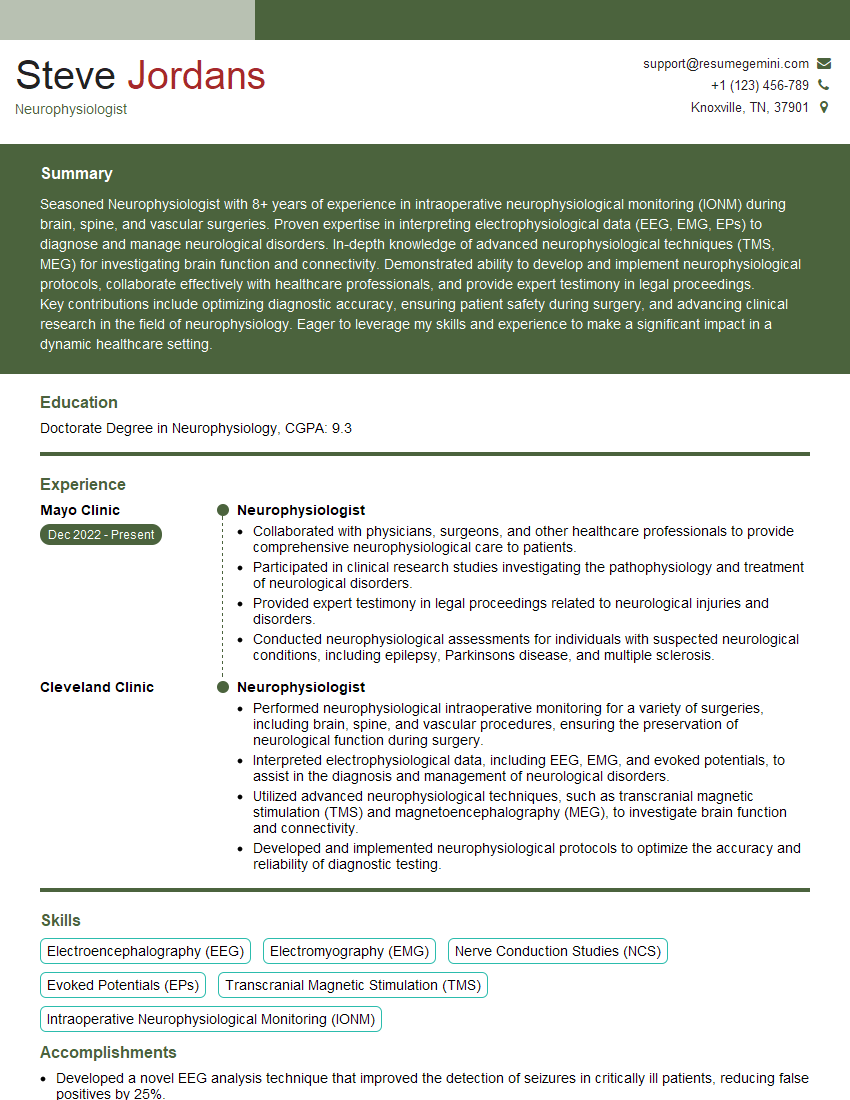
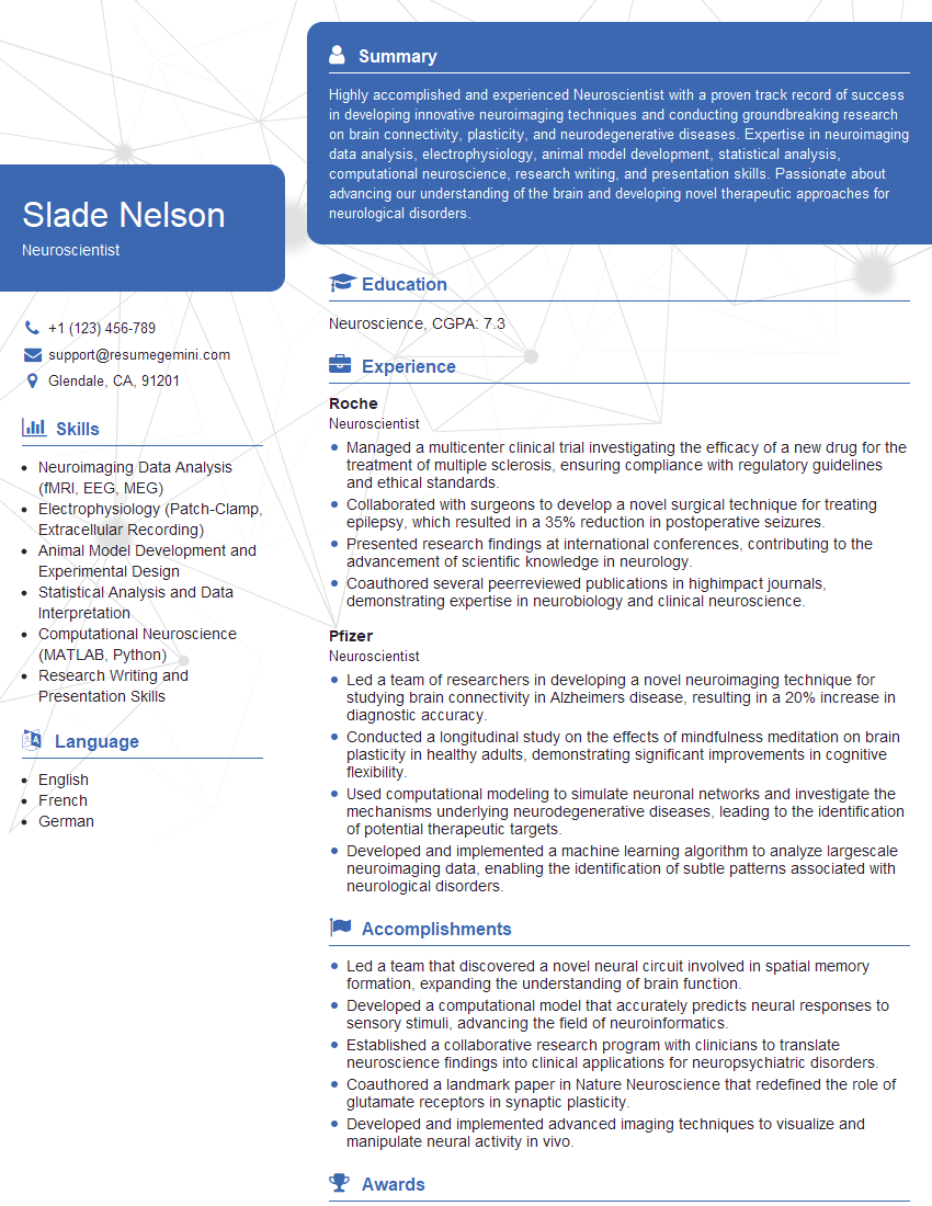
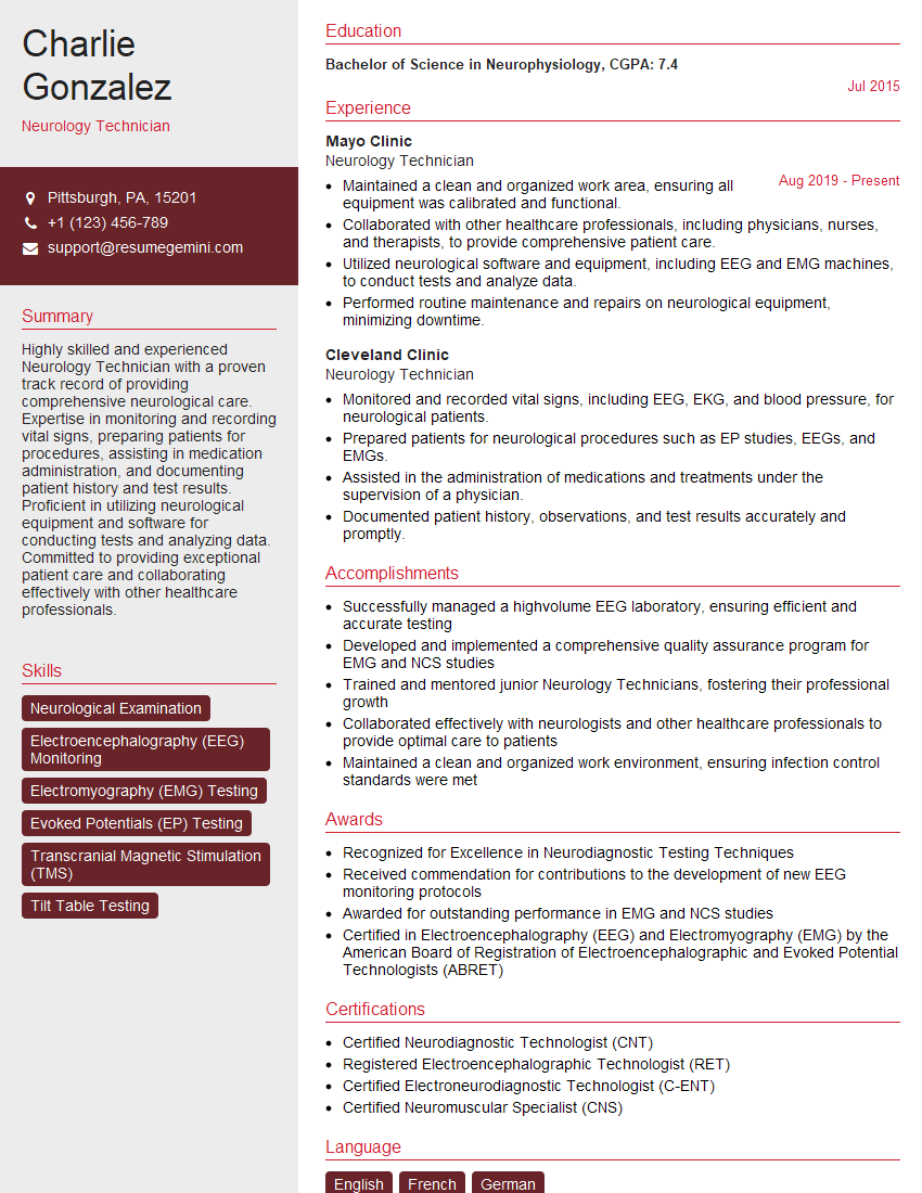
Mastering Neuroanatomy and Neurophysiology is crucial for career advancement in neuroscience research, clinical neurology, and related fields. A strong understanding of these topics demonstrates your expertise and opens doors to exciting opportunities. To maximize your job prospects, it’s vital to create an ATS-friendly resume that highlights your skills and experience effectively. ResumeGemini is a trusted resource to help you build a professional and impactful resume. We provide examples of resumes tailored to Neuroanatomy and Neurophysiology to guide you in crafting your own compelling application materials.
Explore more articles
Users Rating of Our Blogs
Share Your Experience
We value your feedback! Please rate our content and share your thoughts (optional).
What Readers Say About Our Blog
Live Rent Free!
https://bit.ly/LiveRentFREE
Interesting Article, I liked the depth of knowledge you’ve shared.
Helpful, thanks for sharing.
Hi, I represent a social media marketing agency and liked your blog
Hi, I represent an SEO company that specialises in getting you AI citations and higher rankings on Google. I’d like to offer you a 100% free SEO audit for your website. Would you be interested?


