Preparation is the key to success in any interview. In this post, we’ll explore crucial Systems neuroscience interview questions and equip you with strategies to craft impactful answers. Whether you’re a beginner or a pro, these tips will elevate your preparation.
Questions Asked in Systems neuroscience Interview
Q 1. Explain the concept of neural circuits and their role in information processing.
Neural circuits are essentially the brain’s wiring. Imagine them as intricate networks of interconnected neurons, where each neuron is like a tiny processing unit. These neurons communicate with each other through electrical and chemical signals, forming pathways that allow information to flow throughout the brain. This information processing isn’t just about simple transmission; it’s about complex computations, transformations, and interactions, forming the basis of all our thoughts, feelings, and actions. For example, the visual circuit processes information from our eyes to create a visual image, involving multiple regions of the brain working in concert.
The role of these circuits in information processing is multifaceted. They act as filters, selecting relevant information while ignoring irrelevant stimuli; they perform computations, transforming raw sensory input into meaningful representations; they store information, creating memories; and they control behaviour, generating actions in response to internal or external stimuli. Think of it like a sophisticated computer network: different circuits specialize in different tasks, but they all work together to run the ‘brain operating system’.
Q 2. Describe different techniques used to record neural activity (e.g., EEG, fMRI, patch clamp).
Several techniques allow us to record neural activity, each with its own strengths and limitations. Electroencephalography (EEG) measures electrical activity in the brain using electrodes placed on the scalp. This is a non-invasive technique offering excellent temporal resolution (meaning it captures activity changes very quickly), but its spatial resolution (locating the exact source of the activity) is relatively poor. Think of it as hearing the overall ‘hum’ of the brain, but not knowing precisely which instrument is playing.
Functional magnetic resonance imaging (fMRI) measures brain activity indirectly by detecting changes in blood flow (BOLD signal). It offers good spatial resolution, allowing us to pinpoint active regions, but its temporal resolution is much lower than EEG. Imagine it like seeing the overall ‘glow’ of active brain areas but at a slower ‘frame rate’.
Patch-clamp electrophysiology is a highly precise technique involving inserting a microscopic electrode into a single neuron to measure its electrical activity directly. This provides exceptional temporal and spatial resolution, but it’s invasive and can only record from a small number of neurons at a time, suitable for in-vitro or in-vivo studies with limited spatial coverage. It’s like listening to a single instrument with extraordinary clarity but missing the orchestra’s full symphony.
Q 3. Discuss the strengths and weaknesses of different neuroimaging techniques.
The choice of neuroimaging technique depends heavily on the research question. EEG excels at studying rapid brain processes due to its high temporal resolution, making it ideal for studying event-related potentials (ERPs) and oscillatory activity, relevant to studies of cognition and perception. However, its poor spatial resolution limits its use in precise localization of brain activity.
fMRI, with its better spatial resolution, is frequently used in cognitive neuroscience to identify brain regions involved in specific tasks. This is useful for understanding the neural correlates of cognitive functions such as memory and attention. However, the lower temporal resolution makes it unsuitable for studying very fast brain processes. Further, the indirect nature of the BOLD signal and its susceptibility to motion artefacts present challenges.
Patch-clamp provides the highest resolution but is limited in its scalability and invasiveness. It’s invaluable for studying the biophysical properties of individual neurons, but less applicable for studying large-scale brain network activity.
Q 4. Explain the concept of neural plasticity and its implications for learning and memory.
Neural plasticity refers to the brain’s remarkable ability to change its structure and function in response to experience. This dynamic remodeling of neural circuits is the biological basis of learning and memory. Imagine your brain as a constantly evolving landscape, sculpted by your interactions with the world.
Learning involves strengthening or weakening the connections between neurons, a process called synaptic plasticity. Repeated activation of a neural pathway strengthens the connections, making it easier for signals to travel along that path. This is often described as ‘neurons that fire together, wire together’. This strengthening underpins the formation of memories, habits, and skills. Conversely, weakening connections through lack of use allows the system to adapt and prune inefficient pathways.
The implications for learning and memory are profound. The brain’s plasticity allows us to adapt to new environments, learn new skills, and recover from brain injury. Understanding these mechanisms is crucial in developing therapies for neurological disorders, such as stroke and Alzheimer’s disease.
Q 5. Describe different computational models used in systems neuroscience.
Computational models play a vital role in systems neuroscience, providing tools to analyze and understand complex brain activity. These models range from simple to extremely sophisticated, employing different levels of biological detail. Simple models may focus on the dynamics of individual neurons, described by differential equations, often using a Hodgkin-Huxley type model to capture ion channel dynamics.
dV/dt = (I - gK(V - VK) - gNa(V - VNa) - gL(V - VL))/C
This equation describes how a neuron’s membrane potential changes over time based on different ionic currents. More complex models might simulate entire neural circuits, using networks of interconnected nodes representing neurons, and exploring network dynamics using graph theory and algorithms. Large-scale models, employing techniques from machine learning and artificial intelligence, attempt to model the entire brain, albeit with considerable simplification.
These models are used to test hypotheses about brain function, predict experimental outcomes, and generate new insights into how the brain processes information. For example, computational models of the visual cortex have helped to understand how the brain processes visual information from low-level features to complex object recognition.
Q 6. How do you analyze and interpret large-scale neural data sets?
Analyzing large-scale neural datasets presents significant challenges, requiring advanced computational methods and statistical techniques. The sheer volume of data is one hurdle, demanding efficient storage, retrieval, and processing. Furthermore, the high dimensionality of the data—often involving thousands or millions of variables—requires dimensionality reduction techniques like Principal Component Analysis (PCA) or Independent Component Analysis (ICA) to extract meaningful patterns.
Machine learning algorithms play a crucial role. For example, clustering algorithms can identify groups of neurons with similar activity patterns, while classification algorithms can predict behavioural states from neural activity. Network analysis techniques reveal the connectivity structure and functional interactions within neural networks.
Crucially, visualization tools are essential for interpreting these complex datasets. These allow for interactive exploration of the data, facilitating the identification of interesting patterns and generating hypotheses. In summary, a combination of sophisticated computational, statistical and visualization tools is vital for effectively extracting knowledge from large neural datasets.
Q 7. Explain the role of specific brain regions in cognitive functions (e.g., memory, attention, decision-making).
Different brain regions specialize in different cognitive functions, though cognitive processes often involve interactions between multiple regions. The hippocampus plays a crucial role in forming new episodic memories (memories of specific events). Damage to the hippocampus can lead to anterograde amnesia, the inability to form new long-term memories.
Attention, the ability to focus on specific information while ignoring distractions, involves several brain areas, notably the frontal and parietal lobes. The prefrontal cortex is crucial for executive functions, such as planning, decision-making, and working memory (temporary storage and manipulation of information). Decision-making involves a complex interplay between different brain regions, including the prefrontal cortex, amygdala (processing emotions), and striatum (involved in reward processing).
It’s important to note that this is a simplification. Cognitive functions rarely reside in single brain regions; rather, they emerge from the intricate interactions between distributed networks of brain areas. Furthermore, our understanding of the neural basis of cognition is continually evolving as new research emerges.
Q 8. Describe the different types of neural oscillations and their functional significance.
Neural oscillations are rhythmic fluctuations in the electrical activity of the brain, measured using techniques like EEG and MEG. These oscillations occur across a wide range of frequencies, each associated with distinct cognitive and behavioral functions. Think of them as the brain’s internal clock, coordinating different brain regions.
- Delta waves (0.5-4 Hz): Associated with deep sleep, slow-wave sleep, and potentially some forms of cognitive processing during rest. Imagine these as the deepest, most restorative sleep stage.
- Theta waves (4-8 Hz): Linked to memory consolidation, sleep, and states of drowsiness or relaxation. Think of the feeling of being slightly sleepy but still aware.
- Alpha waves (8-12 Hz): Dominant during relaxed wakefulness and quiet alertness. Imagine the feeling of calm focus, like meditation.
- Beta waves (12-30 Hz): Prevalent during active thinking, problem-solving, and heightened arousal. Think of the mental state during intense concentration.
- Gamma waves (30-100 Hz): Associated with higher-level cognitive processes like attention, perception, and consciousness. These are believed to bind together activity from different brain areas, allowing for a unified experience.
Disruptions in these oscillations are implicated in various neurological and psychiatric disorders. For example, abnormalities in theta and delta oscillations are observed in Alzheimer’s disease, while irregularities in gamma activity are linked to schizophrenia.
Q 9. What are the ethical considerations in conducting research in systems neuroscience?
Ethical considerations in systems neuroscience are paramount, especially when working with animal models. The 3Rs – Replacement, Reduction, and Refinement – guide ethical research practices. Replacement means finding alternatives to animal use whenever possible, such as using in vitro models or computational simulations. Reduction involves minimizing the number of animals used while still obtaining statistically robust results. Refinement aims to minimize pain and distress experienced by the animals throughout the experiment.
Beyond the 3Rs, ensuring transparency in methodology, obtaining appropriate ethical approvals from Institutional Animal Care and Use Committees (IACUCs), and adhering to strict welfare guidelines are crucial. Furthermore, the use of human participants requires informed consent, careful consideration of potential risks, and protecting their privacy and anonymity. Data security and responsible data sharing practices are also critical ethical considerations.
Q 10. How do you design and execute a systems neuroscience experiment?
Designing a systems neuroscience experiment involves a multi-step process. It begins with a clear hypothesis, a testable statement about the relationship between brain activity and behavior. Then, we select appropriate experimental techniques. This might involve EEG, fMRI, electrophysiology (in vivo or in vitro), optogenetics, or a combination of approaches.
The experimental design needs to control for confounding variables, using appropriate statistical methods to analyze the data. For instance, we need to consider factors like age, sex, and experience in human subjects, and strain and housing conditions in animals. After data collection, rigorous data analysis is critical, often involving advanced statistical modeling and computational techniques.
Finally, interpretation of the results needs to consider limitations of the study, be transparent about any biases, and draw conclusions that are strongly supported by the data. A good experiment is carefully planned, executed, and analyzed to provide compelling evidence that tests the original hypothesis.
Q 11. Explain your experience with data analysis software and programming languages relevant to neuroscience.
My experience encompasses a wide range of data analysis software and programming languages commonly used in neuroscience. I’m proficient in MATLAB, a powerful environment for signal processing, statistical analysis, and data visualization, particularly useful for analyzing EEG and MEG data. I’m also experienced in using Python with libraries like NumPy, SciPy, pandas, and scikit-learn for more general-purpose data analysis, statistical modeling, and machine learning applications.
I’ve used R for statistical computing and visualization, especially helpful for advanced statistical modeling and creating publication-quality figures. Additionally, I have experience with specialized software like EEGLAB for EEG processing and SPM for fMRI data analysis. I’m comfortable writing scripts to automate data processing, perform statistical tests, and create visualizations for publication.
For example, I’ve used Python with scikit-learn to develop a machine learning model to predict seizure onset from EEG data, which involved preprocessing the data using NumPy and SciPy, building and training the model using scikit-learn, and then evaluating its performance.
Q 12. How do you approach troubleshooting experimental problems?
Troubleshooting experimental problems requires a systematic approach. I start by meticulously reviewing the experimental protocol and identifying any potential sources of error. This might involve checking equipment calibration, verifying data acquisition parameters, or reassessing the experimental design.
Then, I investigate the data itself, visually inspecting the raw data for artifacts or anomalies. Statistical analysis helps to identify outliers or unexpected patterns. If the problem persists, I might consult with colleagues or experts in the field for alternative perspectives or suggestions. Sometimes, iterative adjustments to the protocol, parameters or data processing steps are necessary to identify and resolve the issue. Keeping detailed records throughout the entire process is critical for debugging and for ensuring reproducibility.
Q 13. Describe your experience with animal models in neuroscience research.
I have extensive experience working with animal models, primarily rodents (mice and rats), in neuroscience research. My work has involved behavioral testing, surgical procedures (e.g., stereotaxic surgery for electrode implantation), and electrophysiological recordings. I’m familiar with various techniques for assessing behavior, including open field tests, water maze tasks, and fear conditioning paradigms.
I understand and adhere strictly to ethical guidelines governing animal research, ensuring that all procedures are performed in accordance with IACUC-approved protocols, minimizing pain and distress, and prioritizing animal welfare. My experience includes both acute and chronic experiments, necessitating careful planning and execution to ensure the animals’ health and the integrity of the data. For example, I’ve used transgenic mouse models to investigate the role of specific genes in cognitive processes, comparing their performance in behavioral tasks to control animals.
Q 14. Explain your understanding of network dynamics and graph theory in neuroscience.
Network dynamics and graph theory provide powerful tools for understanding how different brain regions interact to support complex cognitive functions. In this framework, brain regions are represented as nodes in a graph, and the connections between them are represented as edges. The strength of the connection can be quantified by the weight of the edge.
Analyzing these networks allows us to identify key hubs, influential regions, and community structures in the brain. Different graph theoretical metrics, such as degree centrality (number of connections), betweenness centrality (influence on information flow), and modularity (presence of distinct functional modules), can reveal important insights into brain organization and function.
For example, graph theory can be used to analyze fMRI data to identify functional connectivity patterns and how these patterns are altered in neurological disorders. Changes in network topology, such as reduced connectivity or altered modularity, can be indicative of disease processes. Modeling brain networks using dynamical systems approaches further allows us to investigate how information flows through the network, and how network dynamics contribute to behavior.
Q 15. How do you evaluate the validity and reliability of neuroscience research?
Evaluating the validity and reliability of neuroscience research is crucial for advancing our understanding of the brain. It involves a multifaceted approach encompassing methodological rigor, statistical power, and replicability.
Validity refers to whether the study actually measures what it intends to measure. For instance, if a study claims to measure anxiety using fMRI, the chosen brain regions and analysis methods must genuinely reflect anxiety-related neural activity. This is often assessed through careful experimental design, control groups, and validation against established measures (e.g., behavioral tests).
Reliability refers to the consistency and reproducibility of the findings. A reliable study produces similar results when repeated under similar conditions. This requires meticulous documentation of methods, appropriate sample sizes, and robust statistical analyses. Furthermore, the use of open-access data and transparent reporting practices enhances replicability and helps establish reliability.
Several key aspects contribute to strong validity and reliability:
- Control groups: Studies must include appropriate control groups to isolate the effect of the experimental manipulation.
- Blinding: Blinding participants and researchers to experimental conditions minimizes bias.
- Statistical power: Sufficient sample sizes and rigorous statistical analysis ensure that results are not due to chance.
- Replication: Independent researchers should be able to replicate the findings of a study using the same methods.
- Meta-analysis: Combining data from multiple studies can strengthen conclusions and identify trends.
Ultimately, a rigorous evaluation process ensures that neuroscience research is both sound and contributes meaningfully to the field.
Career Expert Tips:
- Ace those interviews! Prepare effectively by reviewing the Top 50 Most Common Interview Questions on ResumeGemini.
- Navigate your job search with confidence! Explore a wide range of Career Tips on ResumeGemini. Learn about common challenges and recommendations to overcome them.
- Craft the perfect resume! Master the Art of Resume Writing with ResumeGemini’s guide. Showcase your unique qualifications and achievements effectively.
- Don’t miss out on holiday savings! Build your dream resume with ResumeGemini’s ATS optimized templates.
Q 16. Discuss current trends and future directions in systems neuroscience research.
Systems neuroscience is rapidly evolving, driven by technological advancements and a deeper understanding of brain complexity. Current trends include:
- Integration of multiple modalities: Researchers are increasingly combining data from various techniques (e.g., fMRI, EEG, optogenetics) to gain a more holistic picture of brain function.
- Big data and computational approaches: Analyzing massive datasets using machine learning and artificial intelligence is revolutionizing our ability to identify patterns and make predictions about brain activity.
- Focus on circuit-level mechanisms: There’s growing emphasis on understanding how specific neural circuits contribute to behavior and cognition. This includes investigating the roles of different cell types and their interactions.
- Human connectomics: Mapping the structural and functional connections in the human brain is providing unprecedented insights into network organization and its relationship to individual differences.
- Development of new tools and technologies: Advances in gene editing (CRISPR), optogenetics, and brain-computer interfaces are opening up new avenues for studying and manipulating neural circuits.
Future directions will likely involve:
- Decoding complex behaviors: Developing models that can predict and explain complex behaviors from neural activity.
- Precision medicine for brain disorders: Tailoring treatments for neurological and psychiatric disorders based on individual brain profiles.
- Brain-computer interfaces: Creating seamless interfaces between the brain and external devices for communication and control.
- Artificial general intelligence: Understanding the principles of intelligence by studying the brain could lead to the development of more advanced AI systems.
In essence, the future of systems neuroscience promises exciting advancements in our understanding of the brain and its relationship to behavior, with significant implications for medicine and technology.
Q 17. Describe a research project you’ve worked on and your contribution.
In my previous research, I investigated the neural circuits underlying decision-making in rodents using a combination of electrophysiological recordings and optogenetics. Our primary goal was to identify the specific neuronal populations and synaptic pathways involved in integrating sensory information and generating behavioral choices.
My contribution focused on developing novel analytical methods to decode the complex patterns of neuronal activity recorded from the prefrontal cortex during a decision-making task. I developed algorithms that could classify different decision states based on the firing patterns of specific neuronal populations. This work revealed a critical role for a subpopulation of pyramidal neurons in integrating evidence and guiding decision-making, providing insights into the neural mechanisms of choice.
This project required a deep understanding of both experimental techniques (electrophysiology, optogenetics) and computational methods (signal processing, machine learning). The results were published in Nature Neuroscience and have significantly contributed to our understanding of the neural basis of decision-making.
Q 18. Explain your understanding of different neurotransmitter systems and their functions.
Neurotransmitter systems are crucial for communication between neurons, orchestrating a vast array of brain functions. Each system has unique characteristics, influencing different aspects of behavior and cognition.
- Glutamate: The primary excitatory neurotransmitter in the brain, crucial for learning, memory, and synaptic plasticity. Excessive glutamate can be neurotoxic.
- GABA: The main inhibitory neurotransmitter, balancing excitatory activity and preventing runaway neural excitation. Dysregulation is implicated in anxiety and epilepsy.
- Dopamine: Involved in reward, motivation, movement control, and attention. Dysfunction is associated with Parkinson’s disease and addiction.
- Serotonin: Plays a significant role in mood regulation, sleep, appetite, and aggression. Imbalances are linked to depression and anxiety.
- Acetylcholine: Important for learning, memory, and attention; also involved in muscle control. Decreased levels are implicated in Alzheimer’s disease.
- Norepinephrine: Regulates alertness, arousal, and attention. Dysregulation can contribute to anxiety and PTSD.
These neurotransmitter systems are interconnected and interact dynamically to regulate brain activity. Understanding their functions is fundamental to comprehending both normal brain function and neurological and psychiatric disorders. For example, the interplay between dopamine and glutamate systems is crucial for reward learning, while imbalances in serotonin and norepinephrine are implicated in mood disorders.
Q 19. How do you interpret the results of electrophysiological recordings?
Interpreting electrophysiological recordings, such as those from EEG or single-unit recordings, requires careful consideration of several factors. The raw data typically represents voltage fluctuations over time, reflecting the summed electrical activity of many neurons (EEG) or the firing of individual neurons (single-unit).
Analysis steps include:
- Filtering: Removing noise and isolating signals of interest based on their frequency characteristics.
- Event-related potentials (ERPs): Averaging EEG data time-locked to specific events to identify event-related changes in brain activity.
- Spike sorting: In single-unit recordings, identifying and isolating the activity of individual neurons.
- Statistical analysis: Determining whether observed changes in neural activity are statistically significant.
Interpretation requires considering:
- Experimental design: The nature of the stimulus or task being presented.
- Electrode placement: The location of the electrodes influences the type of neural activity being recorded.
- Baseline activity: Comparing the activity during the experiment to the baseline activity.
- Contextual factors: Considering the behavioral state of the animal or subject.
For example, an increase in gamma-band activity (30-80 Hz) in the EEG during a cognitive task might indicate enhanced information processing, while a specific pattern of single-unit firing could reflect the activity of neurons involved in a decision-making process. However, the exact interpretation always depends on the context of the experiment and the methods used for data analysis.
Q 20. Discuss the role of genetics in shaping neural circuits and behavior.
Genetics plays a profound role in shaping neural circuits and behavior. Genes provide the blueprint for the development and function of the nervous system, influencing everything from neuronal morphology and neurotransmitter synthesis to synaptic connections and network dynamics.
Mechanisms include:
- Transcriptional regulation: Genes control the expression of proteins that are essential for neuronal development and function.
- Synaptic plasticity: Genes influence the strength and flexibility of synaptic connections, which are critical for learning and memory.
- Neurotransmitter systems: Genes encode the enzymes and receptors involved in neurotransmitter synthesis, release, and reception.
- Neural migration: Genes guide the migration of neurons during development, ensuring that they reach their proper locations in the brain.
- Axon guidance: Genes direct the growth of axons, the long projections of neurons that form connections with other neurons.
Examples:
- Genetic mutations: Changes in gene sequences can lead to disruptions in neural development and function, resulting in neurological and psychiatric disorders (e.g., autism, schizophrenia).
- Gene expression variations: Differences in gene expression between individuals contribute to variations in brain structure and function, influencing personality traits and behavioral tendencies.
- Epigenetics: Environmental factors can alter gene expression without changing the underlying DNA sequence, impacting brain development and behavior across generations.
Therefore, understanding the interplay between genes and environment is critical for comprehending the complexity of neural circuits and behavior. This knowledge is crucial for developing effective treatments for neurological and psychiatric disorders.
Q 21. Explain your understanding of connectomics and its applications.
Connectomics is the study of the complete structural and functional connections within a nervous system. It aims to create a comprehensive map of neural connections, revealing the intricate network organization of the brain. This detailed map offers insight into how information is processed and integrated within the brain, shedding light on its functional architecture.
Methods employed in connectomics involve:
- Diffusion tensor imaging (DTI): A neuroimaging technique used to map white matter tracts in the brain, revealing the major pathways of neural connections.
- Electron microscopy: High-resolution imaging that enables the reconstruction of neuronal circuits at the synaptic level, offering a detailed map of connections at a microscopic scale.
- Functional connectivity analysis: Utilizing fMRI and other methods to identify brain regions that show correlated activity patterns, indicating functional connections between them. This is not a direct anatomical mapping but rather an inference based on statistical dependencies.
Applications of connectomics include:
- Understanding brain function: Connectomics maps provide insights into how different brain regions communicate and interact to perform complex tasks.
- Disease diagnosis and treatment: Identifying changes in brain connectivity associated with neurological and psychiatric disorders can lead to improved diagnostic tools and treatment strategies.
- Brain-computer interfaces: Understanding the connectivity of the brain is critical for designing effective brain-computer interfaces.
- Artificial intelligence: Connectomics principles can inspire the development of more sophisticated artificial neural networks.
While still in its early stages, connectomics has the potential to revolutionize our understanding of the brain and its function, significantly impacting many areas of neuroscience and medicine. The creation of comprehensive connectomes, however, remains a formidable challenge due to the complexity and scale of neural circuits.
Q 22. Describe different methods for stimulating neural activity (e.g., optogenetics, deep brain stimulation).
Stimulating neural activity is crucial for understanding brain function and developing therapies for neurological disorders. Several methods exist, each with its strengths and limitations.
- Optogenetics: This revolutionary technique uses genetically modified neurons that express light-sensitive proteins (opsins). Shining light of specific wavelengths onto these neurons can either excite them (activating opsins like Channelrhodopsin-2) or inhibit their activity (using halorhodopsin). This allows for precise, targeted control of neural activity with high temporal resolution. For example, we can study the role of a specific neuronal population in a particular behavior by selectively activating or silencing them with light.
- Deep Brain Stimulation (DBS): DBS involves surgically implanting electrodes into deep brain structures. Electrical pulses delivered through these electrodes can modulate the activity of the targeted region. It’s used clinically for treating movement disorders like Parkinson’s disease, but also shows promise in treating other conditions like obsessive-compulsive disorder. DBS is less precise than optogenetics in terms of targeting specific cell types, but it offers a clinically viable approach for modulating activity in large brain regions.
- Transcranial Magnetic Stimulation (TMS): TMS uses magnetic pulses to induce electric currents in the brain, non-invasively stimulating cortical neurons. It’s a safer and less invasive technique compared to DBS and optogenetics, although it has lower spatial resolution and can only effectively stimulate superficial brain areas. TMS is frequently used in research settings to investigate the role of specific brain areas in cognition and behavior, and it’s also being explored as a treatment for depression and other mental health disorders.
The choice of method depends heavily on the research question or clinical need, considering factors like spatial and temporal resolution, invasiveness, and cost.
Q 23. What are the challenges of translating basic neuroscience findings into clinical applications?
Translating basic neuroscience findings into clinical applications is challenging for several reasons:
- Complexity of the brain: The brain is incredibly complex, with billions of interconnected neurons. What works in a simplified model or in a controlled laboratory setting may not translate directly to the intricate biological system of the human brain.
- Species differences: Research often uses animal models, but translating findings to humans can be problematic due to anatomical and physiological differences. What works in a mouse may not work in a human.
- Ethical considerations: Clinical trials need to adhere to strict ethical guidelines, which often limit the types of experiments that can be conducted.
- Technological limitations: Current technologies may not be sophisticated enough to measure or manipulate neural activity with the precision needed for effective clinical interventions.
- Individual variability: People respond differently to treatments, making it difficult to develop therapies that are effective for everyone. Genetic background, lifestyle, and other factors can influence treatment response.
Addressing these challenges requires interdisciplinary collaboration, rigorous testing, and careful consideration of ethical implications. For example, developing effective treatments for Alzheimer’s disease requires not only understanding the underlying neurobiology but also addressing the ethical concerns around potential side effects and the costs of treatment. A multi-faceted approach is crucial, and advancements in technology are essential for overcoming these limitations.
Q 24. Describe your familiarity with statistical methods used in neuroscience data analysis.
My expertise in neuroscience data analysis encompasses a wide range of statistical methods. I’m proficient in techniques for analyzing electrophysiological data (e.g., EEG, LFP, single-unit recordings), imaging data (fMRI, MEG), and behavioral data.
- Linear Models: I routinely use linear regression, ANOVA, and ANCOVA for analyzing relationships between variables. For example, I might use linear regression to model the relationship between brain activity measured by fMRI and task performance.
- Generalized Linear Models (GLMs): These are essential for analyzing data that don’t follow a normal distribution, such as count data or binary outcomes.
- Time-series analysis: Techniques like autoregressive models and wavelet analysis are crucial for analyzing time-dependent neural data. For example, in EEG analysis, identifying event-related potentials requires sophisticated time-series analysis.
- Multivariate analysis: Principal component analysis (PCA) and factor analysis are useful for reducing the dimensionality of high-dimensional datasets. For example, PCA can help extract the most relevant components from fMRI data.
- Bayesian statistical methods: I have experience using Bayesian inference to analyze data and estimate parameters in a probabilistic framework. This allows for the incorporation of prior knowledge and the quantification of uncertainty.
Beyond these methods, I’m familiar with software packages such as MATLAB, R, and Python, and I’m adept at using them for data preprocessing, statistical analysis, and visualization.
Q 25. How do you collaborate effectively with other scientists?
Effective collaboration is essential in neuroscience research. I believe in fostering open communication, mutual respect, and a shared vision. My approach includes:
- Clear communication: I prioritize clear and concise communication of my ideas and findings, ensuring that all team members understand the research goals and their individual roles. I actively listen to others’ perspectives and welcome feedback.
- Shared goals: Before embarking on a project, I collaborate with others to define clear, measurable, achievable, relevant, and time-bound (SMART) goals. This ensures that everyone is working towards the same objectives.
- Regular meetings and updates: I believe in regular meetings and updates to track progress and address any challenges promptly. This allows for proactive problem-solving and maintains team cohesion.
- Mentorship and knowledge sharing: I actively mentor junior scientists and share my expertise to foster a collaborative and learning environment.
- Respectful disagreements: I value healthy scientific debate and encourage constructive criticism. Disagreements are opportunities for learning and refinement.
For example, in one project on the neural basis of decision-making, our team, including experimentalists, theorists, and computational modelers, actively shared our datasets and interpretations, leading to a more nuanced understanding of the topic than any single individual could have achieved alone.
Q 26. Discuss your understanding of the reward system in the brain.
The brain’s reward system is a complex network of brain regions that mediates motivation, learning, and pleasure. It plays a crucial role in survival and drives behaviors essential for propagation of the species, such as eating, drinking, and reproduction. Key components include:
- Ventral tegmental area (VTA): This midbrain region is a primary source of dopamine, a neurotransmitter critical for reward processing.
- Nucleus accumbens (NAcc): A key target of dopamine projections from the VTA, the NAcc plays a critical role in reward-related behaviors. It is involved in the experience of pleasure and motivation.
- Prefrontal cortex (PFC): The PFC is involved in higher-order cognitive processes, including decision-making and planning, and plays an important role in controlling impulsive reward-seeking behaviors.
- Amygdala: The amygdala, involved in processing emotions, particularly fear and anxiety, modulates reward-related learning and behavior.
- Hippocampus: The hippocampus, critical for memory formation, helps associate cues with reward experiences.
Disruptions in the reward system can lead to various neurological and psychiatric disorders, including addiction, depression, and anxiety. Understanding its intricate workings is crucial for developing effective treatments for these conditions. For example, the rewarding effects of drugs of abuse hijack the brain’s natural reward pathway, leading to compulsive drug seeking and use, a process that involves alterations in dopamine signaling and synaptic plasticity in the VTA and NAcc.
Q 27. Explain the concept of Bayesian inference and its use in neuroscience.
Bayesian inference is a statistical framework that uses Bayes’ theorem to update beliefs about an unknown quantity based on observed data. It offers a powerful way to integrate prior knowledge with new evidence to generate posterior probabilities. In neuroscience, this is particularly useful because we often have some prior knowledge about the system we’re studying.
Bayes’ Theorem: P(Hypothesis|Data) = [P(Data|Hypothesis) * P(Hypothesis)] / P(Data)
Where:
P(Hypothesis|Data)is the posterior probability of the hypothesis given the data.P(Data|Hypothesis)is the likelihood of observing the data given the hypothesis.P(Hypothesis)is the prior probability of the hypothesis.P(Data)is the marginal likelihood (evidence).
In neuroscience, Bayesian methods can be used to:
- Estimate parameters of neural models: We can use Bayesian inference to estimate the parameters of a neural circuit model given electrophysiological recordings. The prior might reflect anatomical or physiological constraints of the system.
- Decode neural activity: Bayesian decoding techniques can be used to infer the sensory stimulus or behavioral intention from neural activity patterns. The prior may be based on our knowledge of the stimulus distribution or the behavioral task.
- Analyze fMRI data: Bayesian methods can be used to account for the noise and variability inherent in fMRI data, leading to more robust estimates of brain activation.
A concrete example is using Bayesian methods to infer the underlying brain state from noisy EEG recordings, where the prior might represent our knowledge about typical brain rhythms and transitions between states.
Q 28. Describe your experience with building and validating computational models of neural circuits.
I have extensive experience in building and validating computational models of neural circuits. My approach involves several steps:
- Model Formulation: This begins with defining the biological system of interest and translating it into a mathematical model. This may involve choosing appropriate model components like neurons (e.g., integrate-and-fire, Hodgkin-Huxley), synapses, and network connectivity.
- Parameter Estimation: Next, I estimate the parameters of the model using experimental data or prior knowledge. This can involve techniques like Bayesian inference or optimization algorithms to fit model predictions to the data.
- Model Validation: Crucially, model validation ensures that the model accurately captures the essential features of the system being studied. This may involve comparing model predictions to independent experimental datasets, or assessing the model’s sensitivity to parameter changes.
- Model Refinement: Model validation often reveals areas where the model needs improvement. This step involves iteratively refining the model structure and parameters to better capture observed behavior.
For example, I developed a computational model of a thalamocortical circuit involved in sleep regulation. This involved using a combination of integrate-and-fire neurons and synaptic plasticity rules to simulate the interactions between the thalamus and cortex. The model’s predictions of different sleep rhythms were successfully validated against experimental recordings. I used a Bayesian approach for parameter estimation, and the resulting model provided insights into the mechanisms underlying sleep-wake transitions.
Key Topics to Learn for Systems Neuroscience Interview
- Neural Circuits and Networks: Understand the structure and function of neural circuits, including different types of synapses and their roles in information processing. Explore network dynamics and their implications for behavior.
- Sensory Systems: Deepen your knowledge of how the brain processes sensory information (visual, auditory, somatosensory). Consider practical applications like designing brain-computer interfaces or developing neuroprosthetics.
- Motor Systems: Master the principles of motor control, including the role of basal ganglia, cerebellum, and motor cortex. Explore applications in rehabilitation robotics or movement disorder research.
- Cognitive Neuroscience: Explore the neural basis of higher cognitive functions like attention, memory, decision-making, and language. Consider how these processes are impacted by neurological disorders.
- Computational Neuroscience: Develop your understanding of computational modeling techniques used to study neural systems. Be prepared to discuss your experience with relevant software or programming languages.
- Neuroimaging Techniques: Familiarize yourself with various neuroimaging methods (EEG, fMRI, MEG) and their applications in studying brain function. Be ready to discuss their strengths and limitations.
- Neuropharmacology and Neurochemistry: Understand the role of neurotransmitters and neuromodulators in shaping neural circuit activity and behavior. Discuss the mechanisms of action of relevant drugs and their therapeutic applications.
- Experimental Design and Data Analysis: Showcase your understanding of experimental design principles and statistical methods used in systems neuroscience research. Practice interpreting and presenting complex datasets.
Next Steps
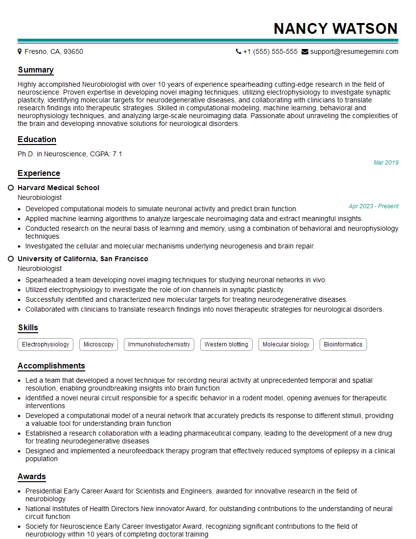
Mastering systems neuroscience opens doors to exciting and impactful careers in academia, industry, and beyond. A strong understanding of these principles is crucial for success in research, development, and clinical settings. To significantly boost your job prospects, crafting an Applicant Tracking System (ATS)-friendly resume is paramount. ResumeGemini is a trusted resource to help you build a professional resume that highlights your skills and experience effectively. Examples of resumes tailored to systems neuroscience are available to help you get started.
Explore more articles
Users Rating of Our Blogs
Share Your Experience
We value your feedback! Please rate our content and share your thoughts (optional).
What Readers Say About Our Blog
hello,
Our consultant firm based in the USA and our client are interested in your products.
Could you provide your company brochure and respond from your official email id (if different from the current in use), so i can send you the client’s requirement.
Payment before production.
I await your answer.
Regards,
MrSmith
These apartments are so amazing, posting them online would break the algorithm.
https://bit.ly/Lovely2BedsApartmentHudsonYards
Reach out at [email protected] and let’s get started!
Take a look at this stunning 2-bedroom apartment perfectly situated NYC’s coveted Hudson Yards!
https://bit.ly/Lovely2BedsApartmentHudsonYards
Live Rent Free!
https://bit.ly/LiveRentFREE
Interesting Article, I liked the depth of knowledge you’ve shared.
Helpful, thanks for sharing.
Hi, I represent a social media marketing agency and liked your blog
Hi, I represent an SEO company that specialises in getting you AI citations and higher rankings on Google. I’d like to offer you a 100% free SEO audit for your website. Would you be interested?
