Cracking a skill-specific interview, like one for Bladder Surgery, requires understanding the nuances of the role. In this blog, we present the questions you’re most likely to encounter, along with insights into how to answer them effectively. Let’s ensure you’re ready to make a strong impression.
Questions Asked in Bladder Surgery Interview
Q 1. Describe the different surgical approaches for bladder cancer resection.
Bladder cancer resection surgery aims to remove cancerous tissue from the bladder. The approach depends on the tumor’s size, location, and stage, as well as the patient’s overall health. Several techniques exist:
- Transurethral Resection of Bladder Tumor (TURBT): This minimally invasive procedure uses a thin, telescope-like instrument inserted through the urethra to remove the tumor. It’s suitable for superficial bladder cancers. Imagine it like using a tiny, specialized scalpel to scrape away a small blemish.
- Partial Cystectomy: This involves removing only the cancerous portion of the bladder, preserving as much healthy tissue as possible. Think of it as surgically excising a specific area of the bladder, similar to removing a small, localized growth from another organ.
- Radical Cystectomy: This is a more extensive surgery where the entire bladder, surrounding lymph nodes, and portions of the prostate (in men) or uterus/vagina (in women) are removed. It’s used for more advanced cancers that have infiltrated the bladder wall. This is a more aggressive approach, analogous to removing an entire infected limb to prevent further spread.
The choice of surgical approach is crucial and depends on a careful assessment of the tumor and the patient’s condition. We always aim for the least invasive surgery that effectively addresses the cancer.
Q 2. Explain the principles of radical cystectomy.
Radical cystectomy is a complex procedure with several key principles: complete tumor removal, adequate lymph node dissection, and urinary diversion. Let’s break it down:
- Complete Tumor Removal: The entire bladder, along with a margin of healthy tissue surrounding the tumor, is removed to ensure no cancer cells are left behind. This is paramount to preventing recurrence.
- Lymph Node Dissection: Surrounding lymph nodes are removed and examined under a microscope to check for cancer spread. This helps in staging the cancer and guiding further treatment. Think of it as investigating the ‘neighborhood’ surrounding the tumor to assess the spread.
- Urinary Diversion: Since the bladder is removed, a new way to drain urine needs to be created. Options include an ileal conduit (using a segment of the small intestine to create a stoma), a neobladder (creating a new bladder from intestinal tissue), or urinary diversion to the ureters. The method chosen is highly individualized based on patient factors and preferences.
The precise steps involved vary depending on the specific circumstances. Pre-operative planning, meticulous surgical technique, and close post-operative monitoring are essential for a successful outcome. We meticulously plan each surgery to minimize risks and optimize results for each patient.
Q 3. What are the indications and contraindications for bladder augmentation?
Bladder augmentation is a surgical procedure to increase the bladder’s capacity. It’s primarily used in patients with a neurogenic bladder (reduced bladder capacity due to neurological conditions) or those with a very small bladder.
- Indications: Frequent urination, urinary urgency, incontinence, low bladder capacity causing frequent infections or complications related to chronic bladder emptying issues.
- Contraindications: Active infection, severe bladder inflammation, certain systemic illnesses, and the presence of certain cancers can prevent augmentation. The decision is made on a case-by-case basis after a thorough evaluation of the patient’s condition.
A common example is a child born with a very small bladder; augmentation can provide relief from significant bladder discomfort and urinary symptoms. For instance, we can utilize bowel segments to build up bladder volume. The choice of augmentation material is important and discussed thoroughly with the patient.
Q 4. Discuss the management of urinary incontinence post-prostatectomy.
Urinary incontinence after prostatectomy is a common complication. Management involves a multi-pronged approach:
- Conservative Measures: Pelvic floor exercises (Kegel exercises) to strengthen pelvic muscles, bladder training techniques to regulate voiding, and lifestyle modifications (fluid intake adjustments).
- Medications: Certain medications can help improve bladder control by reducing muscle spasms or increasing urethral resistance.
- Surgical Interventions: If conservative measures fail, surgical options, such as sling procedures, artificial urinary sphincters, or bulking agents, might be considered. These aim to restore urethral sphincter function or provide additional support to the urethra.
The specific management strategy depends on the type and severity of incontinence. We tailor the treatment plan to individual patient needs, often employing a stepwise approach starting with conservative measures and progressing to more invasive interventions as needed. For example, we’ll assess whether incontinence is stress-related, urge-related, or a combination.
Q 5. How do you assess bladder function pre- and post-operatively?
Assessing bladder function involves a comprehensive evaluation before and after surgery. This includes:
- Pre-operative Assessment: Uroflowmetry (measuring urine flow rate), post-void residual (measuring urine left in the bladder after urination), cystometry (measuring bladder pressure and capacity), and imaging studies (ultrasound or CT scan) to assess bladder anatomy and function.
- Post-operative Assessment: Similar tests are repeated to monitor recovery, detect complications, and assess the success of the surgery. Additional tests might include urine cultures to check for infections and blood tests to monitor renal function.
These tests provide crucial information for planning surgery, guiding treatment decisions, and monitoring the patient’s recovery process. For example, a low post-void residual indicates effective bladder emptying after prostatectomy.
Q 6. What are the common complications of bladder surgery?
Bladder surgery, while often successful, carries several potential complications:
- Infection: Urinary tract infections are common, particularly after procedures involving urinary diversion.
- Bleeding: Bleeding can occur during or after surgery, requiring intervention.
- Urinary Incontinence: Damage to nerves or muscles controlling bladder emptying can result in temporary or permanent incontinence.
- Strictures: Narrowing of the urethra or ureter can occur, requiring further treatment.
- Stoma Complications (if applicable): Patients with a urinary diversion might experience complications such as stoma stenosis, prolapse, or parastomal hernia.
- Sexual Dysfunction (in men): Nerve damage during prostatectomy can lead to erectile dysfunction or retrograde ejaculation.
The risk of these complications varies depending on several factors, including the type of surgery performed, the patient’s overall health, and the surgeon’s experience. We strive to minimize these risks through meticulous surgical technique and careful post-operative management.
Q 7. Describe your experience with robotic-assisted bladder surgery.
I have extensive experience with robotic-assisted bladder surgery. Robotics offers several advantages over traditional open surgery, including:
- Enhanced Precision and Dexterity: The robotic arms allow for finer movements and greater precision during complex surgical procedures.
- Minimally Invasive Approach: Smaller incisions lead to less pain, reduced blood loss, faster recovery times, and improved cosmetic outcomes.
- Improved Visualization: High-definition 3D vision provides a superior view of the surgical field, improving accuracy.
In my practice, we routinely utilize robotic-assisted techniques for radical cystectomies, partial cystectomies, and bladder augmentation. For instance, the enhanced precision allows for a more precise lymph node dissection during radical cystectomy, contributing to better oncological outcomes. Robotics have become an integral part of my surgical practice, allowing us to offer our patients the best possible surgical care.
Q 8. How do you manage urinary tract infections in patients undergoing bladder surgery?
Managing urinary tract infections (UTIs) in patients undergoing bladder surgery is crucial for preventing complications and ensuring a successful outcome. Pre-operative screening with urine cultures is essential to identify and treat any existing infection before surgery. Prophylactic antibiotics are often administered before, during, and after the procedure to reduce the risk of post-operative UTIs. Post-operatively, we closely monitor patients for symptoms like fever, burning during urination, or cloudy urine. If a UTI is suspected, a urine culture is performed to identify the causative organism and guide antibiotic therapy. The choice of antibiotic depends on the specific bacteria identified and its antibiotic sensitivity profile. We also emphasize patient education on hygiene practices to prevent future infections.
For example, a patient presenting with symptoms suggestive of a UTI before a scheduled cystectomy would receive urine culture and sensitivity testing. Based on the results, appropriate antibiotics would be administered before surgery, during surgery with irrigation, and then a course of oral antibiotics post-operatively. This is crucial to prevent a prolonged infection potentially impacting the surgical outcome.
Q 9. What are the different types of urinary diversions and when would you choose each?
Urinary diversions are surgical procedures that reroute urine flow when the bladder is removed or unable to function normally. Several types exist, each suited for different clinical scenarios:
- Ileal conduit (ileostomy): A segment of the ileum (small intestine) is used to create a conduit that drains urine to a stoma (opening) on the abdomen, requiring a pouch for collection. This is a common choice for patients with bladder cancer requiring cystectomy where preservation of bladder function is not possible.
- Neobladder (orthotopic neobladder): A new bladder is constructed using a segment of the bowel, allowing urine to be stored and emptied normally through the urethra. This offers a more natural way of life but is more technically challenging and requires careful patient selection. For example, patients with good bowel function and who are motivated to learn intermittent self-catheterization are ideal candidates.
- Ureterostomy: The ureters (tubes connecting the kidneys to the bladder) are surgically connected to an opening on the abdomen, creating a stoma that directly drains urine. This is usually a temporary solution or considered for patients who are not suitable for other diversions.
The choice depends on factors like the patient’s overall health, surgical expertise, and the patient’s preferences. A thorough discussion outlining the benefits and limitations of each option is crucial for shared decision-making.
Q 10. Explain the role of neoadjuvant chemotherapy in bladder cancer.
Neoadjuvant chemotherapy in bladder cancer refers to chemotherapy administered before surgery. Its primary goal is to shrink the tumor, making surgery easier and potentially improving the chances of complete removal. This is particularly important for muscle-invasive bladder cancers (stages T2-T4). By reducing the tumor burden, neoadjuvant chemotherapy can potentially decrease the risk of local recurrence and improve survival rates. The specific regimen of chemotherapy is tailored to individual patient factors like overall health, tumour grade, and stage.
For instance, a patient with a T3 bladder cancer might receive several cycles of cisplatin-based chemotherapy before undergoing radical cystectomy. The goal is to shrink the tumor, making the surgical procedure less extensive and improving chances of complete resection, consequently improving overall survival.
Q 11. Describe your approach to managing a patient with recurrent bladder cancer.
Managing recurrent bladder cancer requires a multidisciplinary approach involving urologists, oncologists, and radiologists. The treatment strategy is tailored to several factors, including the location and extent of recurrence, previous treatments received, and the patient’s overall health. Options may include:
- Bacillus Calmette-Guérin (BCG) immunotherapy: This is often used for non-muscle invasive recurrences.
- Transurethral resection of bladder tumor (TURBT): This minimally invasive procedure removes recurrent tumors from the bladder lining.
- Chemotherapy: Systemic chemotherapy may be used for more advanced recurrences, either alone or combined with other treatments.
- Radiation therapy: Radiation can be employed as a primary treatment or combined with chemotherapy.
- Cystectomy: In cases of widespread recurrence, cystectomy may be necessary.
Each case is unique and requires careful consideration of the risk-benefit profile of various treatment strategies, always keeping the patient’s quality of life as a priority.
Q 12. What are the latest advancements in bladder cancer treatment?
Bladder cancer treatment is constantly evolving. Recent advancements include:
- Targeted therapies: Drugs that specifically target cancer cells, minimizing damage to healthy tissue, are becoming increasingly prevalent, especially for specific genetic mutations.
- Immunotherapy: Utilizing the body’s immune system to fight cancer cells is showing promising results in advanced bladder cancer, enhancing the response to chemotherapy and improving survival.
- Improved surgical techniques: Minimally invasive approaches like robotic-assisted surgery are reducing complications and recovery time. Robotic-assisted radical cystectomy allows for greater precision and dexterity in complex cases, leading to improved oncological outcomes and faster recovery.
- Advanced imaging techniques: Improved imaging modalities help in early detection and more precise staging of bladder cancer, allowing for more personalized treatment plans. This results in better patient stratification and more accurately targeted therapy.
These advancements offer hope for improved outcomes and a better quality of life for bladder cancer patients.
Q 13. How do you counsel patients about the risks and benefits of bladder surgery?
Counseling patients about bladder surgery involves a sensitive and thorough discussion of the risks and benefits. This is a collaborative process ensuring informed consent. We discuss the potential benefits, such as cure or improved quality of life, alongside potential complications, including infection, bleeding, and urinary or bowel dysfunction. The potential need for urinary diversion and its implications are explained in detail, including stoma care and lifestyle adjustments. Realistic expectations are set regarding recovery time and potential long-term effects. We emphasize the importance of patient participation in decision-making and encourage them to ask questions and express their concerns. Open communication and shared decision-making are paramount throughout the process. For example, I often use diagrams and visual aids to explain the surgical procedure and the impact on urinary function, allowing the patient to actively participate in the decision-making process.
Q 14. Explain your experience with minimally invasive surgical techniques for bladder surgery.
My experience with minimally invasive surgical techniques, particularly robotic-assisted surgery, has been extensive and rewarding. Robotic-assisted radical cystectomy (RARC) offers several advantages over open surgery, including smaller incisions, reduced blood loss, less pain, and shorter hospital stays. The enhanced precision and dexterity of the robotic arms allow for more complex dissections with improved oncological outcomes, particularly when preserving neurovascular bundles for better erectile function in men. While RARC requires a steep learning curve and specialized equipment, the benefits for carefully selected patients are substantial. The improved visualization and precision are particularly valuable in cases with advanced disease or difficult anatomy. In my practice, I routinely employ RARC for suitable candidates and have observed consistently positive results in terms of patient recovery and oncologic outcomes. I also have significant experience with laparoscopic techniques for less extensive bladder surgeries.
Q 15. What is your experience with managing complications such as fistula formation?
Fistula formation, an abnormal connection between the bladder and another organ (like the vagina or rectum), is a serious complication following bladder surgery. Management depends heavily on the type and location of the fistula, as well as the patient’s overall health.
My approach involves a thorough evaluation using imaging techniques like cystoscopy and MRI to precisely locate and characterize the fistula. Conservative management, initially, might include placement of a urinary diversion (like a suprapubic catheter) to allow the fistula to heal spontaneously. This gives the body a chance to close the tract naturally. However, if conservative management fails, surgical repair is necessary. This repair can range from simple closure techniques to more complex reconstructive procedures, depending on the severity. Post-operative care includes close monitoring for recurrence and management of any associated infections.
For example, I recently managed a patient who developed a vesicovaginal fistula after a hysterectomy. Initial conservative management with a catheter failed. Therefore, we proceeded with a transvaginal repair which successfully closed the fistula. Post-operative follow-up was key to ensure complete healing and prevent recurrence.
Career Expert Tips:
- Ace those interviews! Prepare effectively by reviewing the Top 50 Most Common Interview Questions on ResumeGemini.
- Navigate your job search with confidence! Explore a wide range of Career Tips on ResumeGemini. Learn about common challenges and recommendations to overcome them.
- Craft the perfect resume! Master the Art of Resume Writing with ResumeGemini’s guide. Showcase your unique qualifications and achievements effectively.
- Don’t miss out on holiday savings! Build your dream resume with ResumeGemini’s ATS optimized templates.
Q 16. Describe your approach to managing patients with neurogenic bladder.
Neurogenic bladder, where nerve damage impacts bladder function, requires a multi-faceted approach tailored to the individual patient. The underlying neurological condition significantly influences treatment. A complete neurological examination is essential to identify the cause (e.g., spinal cord injury, multiple sclerosis).
Management often involves a combination of strategies. This may include intermittent catheterization (self-catheterization or caregiver-assisted), medication to manage bladder spasms or increase bladder emptying, and surgical options such as augmentation cystoplasty (increasing bladder capacity) or urinary diversion. Regular urodynamic studies are crucial to monitor bladder function and adjust treatment as needed.
Imagine a patient with multiple sclerosis experiencing urinary retention. We would conduct urodynamic studies to assess bladder pressures and emptying capabilities. We might start with medication to help the bladder contract more effectively. If that isn’t sufficient, we may consider intermittent catheterization. In severe cases where the bladder is significantly damaged, surgical intervention such as augmentation cystoplasty would be considered.
Q 17. How do you diagnose and manage interstitial cystitis?
Interstitial cystitis (IC), also known as bladder pain syndrome, is a chronic condition causing bladder pain and urgency. Diagnosis is challenging as there’s no single definitive test. It’s primarily a diagnosis of exclusion, meaning other causes of bladder problems need to be ruled out.
Diagnosis begins with a detailed history and physical examination, focusing on symptoms and ruling out infections. Cystoscopy (direct visualization of the bladder lining) is often performed to assess for characteristic changes, though these aren’t always present. Urodynamic studies assess bladder function and pressure. A bladder biopsy might be considered, though it is not always diagnostic.
Management is individualized and often focuses on pain relief and symptom control. This can involve medications such as amitriptyline or pentosan polysulfate, bladder instillations (directly injecting medication into the bladder), physical therapy, and dietary modifications. In severe cases, more invasive procedures such as nerve stimulation or surgery might be considered.
For example, a patient presenting with chronic pelvic pain and frequent urination would undergo a thorough evaluation including urine culture (to rule out infection), cystoscopy, and urodynamic testing. Depending on the findings, we would then develop a personalized treatment plan, potentially starting with conservative measures like medication and lifestyle changes before progressing to more advanced options.
Q 18. What is your experience with different types of bladder slings?
Bladder slings are surgical procedures used to treat stress urinary incontinence (SUI), where urine leaks with activities like coughing or sneezing. Several types exist, each with advantages and disadvantages.
I have extensive experience with different sling types, including retropubic slings (TVT, TOT, and tension-free vaginal tape), transobturator slings, and single-incision slings (mini-slings). The choice of sling depends on factors such as the patient’s anatomy, previous surgeries, and surgeon preference.
Retropubic slings are commonly used and generally have high success rates, though they carry a slightly higher risk of complications such as bladder injury. Transobturator slings offer a less invasive approach with a shorter operative time, but they have a slightly higher rate of sling erosion. Single-incision slings are minimally invasive, but may not be suitable for all patients. Post-operative management includes careful monitoring for complications such as infection, bleeding, and urinary retention.
For instance, a patient with SUI following childbirth might be a good candidate for a tension-free vaginal tape (TVT) sling procedure due to its effectiveness and relatively low complication rate. However, a patient with a history of pelvic surgeries might be better suited to a less invasive transobturator approach to minimize the risk of complications.
Q 19. Describe your experience with Botox injections for overactive bladder.
Botox injections into the bladder detrusor muscle are a minimally invasive treatment option for overactive bladder (OAB), characterized by urinary urgency, frequency, and urge incontinence. Botox temporarily weakens the bladder muscle, reducing its contractions and improving symptoms.
My experience with Botox injections includes patient selection, injection technique, and post-injection management. Careful patient selection is crucial; Botox is not suitable for all patients with OAB. For example, patients with urinary retention or neurogenic bladder are not ideal candidates. The injection procedure itself is relatively straightforward but requires precise targeting of the detrusor muscle under cystoscopic guidance.
Post-injection, patients are monitored for urinary retention and other potential side effects like urinary tract infections. The effects of Botox are typically temporary, lasting for several months, requiring repeat injections. I often use a combination of Botox and behavioral modification techniques for improved long-term outcomes.
A patient with severe OAB symptoms refractory to oral medications may benefit from Botox injections. However, before this treatment is considered, we would first exhaust conservative management options like bladder training and medication. If these options fail, Botox injections would be considered as an intermediate step before evaluating the need for more invasive surgical options.
Q 20. Explain the role of imaging (e.g., CT, MRI, cystoscopy) in bladder surgery.
Imaging plays a vital role in the diagnosis and management of bladder conditions. Different modalities offer unique insights.
Cystoscopy, a direct visualization of the bladder’s interior using a thin, flexible tube with a camera, is invaluable for detecting bladder tumors, stones, inflammation (like in interstitial cystitis), and assessing the extent of fistulas. CT scans are useful for evaluating the surrounding structures, identifying kidney stones that might be contributing to bladder issues, and assessing for other pelvic abnormalities. MRI provides detailed anatomical imaging, particularly useful for identifying tumors, fistulas, and evaluating the extent of pelvic organ prolapse.
Pre-operatively, imaging helps to plan the surgical approach. For example, a CT scan might reveal the location and size of a bladder stone before a procedure to remove it. Post-operatively, imaging helps monitor healing and detect any complications such as hematomas or abscesses. In a patient with suspected bladder cancer, a cystoscopy coupled with CT or MRI would be used for accurate diagnosis and staging. Each modality provides complementary information to aid in comprehensive diagnosis and treatment planning.
Q 21. How do you manage post-operative pain in patients after bladder surgery?
Post-operative pain management after bladder surgery is crucial for patient comfort and recovery. A multimodal approach is often employed, combining different pain relief methods.
This might include medications such as analgesics (acetaminophen, opioids if necessary), and anti-inflammatories (NSAIDs). Regional anesthesia techniques (e.g., nerve blocks) can provide targeted pain relief. Non-pharmacological methods like heat/ice packs, physiotherapy, and relaxation techniques are also valuable. Regular pain assessments are critical to monitor effectiveness and adjust treatment as needed. Careful consideration should be given to potential side effects, particularly with opioid analgesics, and alternative approaches should be explored whenever possible to minimize the use of potentially addictive medications.
For instance, after a radical cystectomy (removal of the bladder), a patient might receive a combination of intravenous analgesics initially, followed by a transition to oral pain medications as tolerated. Physiotherapy may be introduced to aid in mobility and reduce muscle spasms. Pain management plans are often adjusted based on the individual patient’s response and overall condition, ensuring optimal comfort and a smooth recovery.
Q 22. What are the different types of bladder stones and how are they managed?
Bladder stones, or urolithiasis, are crystalline masses that form within the urinary bladder. They can vary significantly in composition, size, and number. Different types include:
- Calcium stones: The most common type, often composed of calcium oxalate or calcium phosphate. These are usually associated with dietary factors or metabolic disorders.
- Struvite stones: Associated with urinary tract infections (UTIs), particularly those caused by urease-producing bacteria. These stones grow rapidly and can become quite large.
- Uric acid stones: These are less common and often form in individuals with gout or those on certain medications.
- Cystine stones: Relatively rare, these are caused by a genetic disorder leading to increased cystine excretion in the urine.
Management depends on the size, number, and composition of the stones, as well as the patient’s overall health. Small stones may pass spontaneously with increased fluid intake and pain medication. Larger stones, however, often require intervention. This can include:
- Cystoscopy: A minimally invasive procedure where a thin, flexible tube with a camera is inserted into the urethra to visualize and remove stones using a variety of instruments.
- Lithotripsy (shock wave or laser): This breaks up the stones into smaller fragments that can then be passed more easily. Shock wave lithotripsy uses sound waves from outside the body, while laser lithotripsy uses a laser fiber inserted through a cystoscope.
- Open surgery: Reserved for cases where other methods fail, or for very large or complex stones. This involves a larger incision to access and remove the stones directly.
The choice of management strategy is always individualized and involves careful consideration of the patient’s specific situation.
Q 23. Describe your experience with urethral stricture repair.
Urethral stricture repair is a common procedure I perform. A urethral stricture is a narrowing of the urethra, the tube that carries urine from the bladder out of the body. This narrowing can cause difficulty urinating, urinary tract infections, and even kidney damage. The causes are varied, including trauma (e.g., pelvic fractures), infection, or prior surgery.
My approach involves a thorough evaluation, including history, physical exam, and imaging studies (uroflowmetry, urethrography). The choice of surgical technique depends on the location, length, and severity of the stricture. Options include:
- Urethrotomy: A minimally invasive procedure where a small incision is made in the stricture using a specialized instrument passed through the urethra. This is suitable for short, less severe strictures.
- Urethral reconstruction: This involves using a graft (usually buccal mucosa) or flap of tissue to replace the scarred tissue causing the narrowing. This is necessary for longer, more complex strictures. Precise surgical technique is vital here to ensure the graft or flap is well-vascularized and heals properly.
- Urethral dilation: This is a less invasive approach, useful for certain types of strictures and often employed in the post-operative phase. It involves gradually widening the urethra using progressively larger instruments. However, it is important to note that this is often not a durable long-term solution and can lead to recurrence.
Post-operative management involves careful monitoring for complications like infection or re-stricture formation, and often includes urethral dilation. Patient selection and technique optimization are crucial to achieving optimal long-term outcomes.
Q 24. Explain the principles of bladder neck reconstruction.
Bladder neck reconstruction aims to restore normal bladder outlet function. The bladder neck is the area where the bladder connects to the urethra. Problems in this area can lead to urinary incontinence, urinary retention, or both. The principles are based on restoring the normal anatomical and functional relationship between the bladder and urethra, thereby achieving continence or improving voiding function.
The specific technique depends on the underlying cause. Common causes requiring reconstruction include:
- Congenital anomalies: Conditions present from birth like bladder exstrophy.
- Post-prostatectomy incontinence: Following removal of the prostate gland.
- Trauma: Injury to the bladder neck.
- Neurogenic bladder: Bladder dysfunction due to nerve damage.
Techniques range from minimally invasive procedures like endoscopic injection of bulking agents to more complex open surgical procedures involving the use of grafts or flaps. The goal is always to provide sufficient support to the bladder neck and prevent stress urinary incontinence without causing outflow obstruction.
Success depends on careful patient selection, meticulous surgical technique, and post-operative management. I always thoroughly discuss the potential benefits, risks, and alternatives with each patient to ensure informed consent.
Q 25. What is your experience with treating bladder exstrophy?
Bladder exstrophy is a complex congenital anomaly where the bladder is exposed on the lower abdominal wall. It’s a challenging condition requiring a multidisciplinary approach, including urologists, plastic surgeons, and other specialists. My experience involves managing patients from infancy to adulthood.
The primary goal is to close the bladder and reconstruct the abdominal wall. This typically involves a series of surgical procedures, starting in infancy with primary closure of the bladder and often requiring several reconstructive steps throughout childhood. Advanced techniques such as augmentation cystoplasty (bladder enlargement using bowel segments) are sometimes used to increase bladder capacity and improve function.
Long-term follow-up is crucial, as these patients often experience complications like recurrent UTIs, incontinence, and renal dysfunction. It’s vital to provide comprehensive care and support throughout their lives, addressing the functional and cosmetic aspects of the condition. Collaborating closely with other specialists is critical in achieving good outcomes.
Q 26. Describe your approach to managing a patient with a vesicovaginal fistula.
A vesicovaginal fistula (VVF) is an abnormal connection between the bladder and vagina. This leads to continuous or intermittent leakage of urine into the vagina. The causes are varied, ranging from obstetric trauma (most common) to pelvic surgery, radiation therapy, or infection.
My approach involves a thorough evaluation including history, physical exam, and imaging (cystography, MRI). Management typically involves surgical repair, aiming to close the fistula and restore the integrity of the bladder and vaginal walls. The surgical technique is tailored to the specific location, size, and cause of the fistula. It might include simple direct closure, interposition of a graft (e.g., buccal mucosa), or more complex reconstructive procedures. Pre-operative assessment, including bowel preparation, and careful post-operative management, including adequate drainage and infection control, are crucial.
Successful repair relies heavily on meticulous surgical technique and thorough post-operative care. The complexity of the procedure and the patient’s overall health significantly influence the outcome.
Q 27. What is your experience with treating ureterovesical junction obstruction?
Ureterovesical junction (UVJ) obstruction is a blockage at the point where the ureter (tube connecting kidney to bladder) enters the bladder. This can lead to hydronephrosis (swelling of the kidney due to urine backup) and potential kidney damage. The causes are diverse, from congenital abnormalities to acquired conditions like stones or inflammation.
My approach involves a thorough evaluation, including imaging studies (ultrasound, CT scan, or IVP) to assess the degree of obstruction and kidney function. Treatment options range from conservative measures for mild cases to surgical interventions for more severe obstructions. Conservative management may include medications to reduce inflammation or stents to temporarily relieve obstruction. However, surgical intervention is often necessary for long-term resolution.
Surgical options include endoscopic techniques, such as minimally invasive balloon dilation or incision of the obstruction, or open surgery, such as ureteropyeloplasty, which involves reconstructing the UVJ. The selection of surgical technique depends on the individual case and the severity of obstruction.
Q 28. How do you assess and manage patients with urinary retention?
Urinary retention is the inability to completely empty the bladder. It’s a significant problem that can lead to complications such as bladder distension, urinary tract infections, and kidney damage. Causes are multifaceted, from benign prostatic hyperplasia (BPH) and urethral strictures to neurological disorders or medications.
My assessment involves a comprehensive history, physical examination (including palpation of the bladder), and laboratory tests (urinalysis to rule out infection). Imaging studies like ultrasound can measure post-void residual urine volume. The immediate management involves relieving the retention, usually with catheterization. This can be done via urethral catheter or suprapubic catheter.
The long-term management depends on the underlying cause. For BPH, options include medication, minimally invasive procedures (e.g., transurethral resection of the prostate, or TURP), or surgery. Urethral strictures require the procedures discussed previously. Neurogenic bladder management often involves intermittent self-catheterization, medication, or surgical options like bladder augmentation. It’s crucial to determine and address the root cause to prevent recurrence.
Key Topics to Learn for Bladder Surgery Interview
- Bladder Cancer Diagnosis and Staging: Understanding TNM staging, imaging techniques (CT, MRI, cystoscopy), and biopsy interpretation.
- Surgical Techniques for Bladder Cancer: Proficiency in radical cystectomy, partial cystectomy, and urinary diversion procedures (ileal conduit, orthotopic neobladder, etc.). Practical application includes understanding indications, contraindications, and potential complications for each.
- Minimally Invasive Approaches: Knowledge of robotic-assisted radical cystectomy, laparoscopic techniques, and their advantages and limitations compared to open surgery.
- Urinary Reconstruction and Diversion: Deep understanding of various urinary diversion techniques, their associated complications (e.g., strictures, leakages, infections), and management strategies.
- Management of Post-Operative Complications: Addressing potential complications such as bleeding, infection, urinary fistulas, and bowel injury; problem-solving approaches focusing on early detection and intervention.
- Functional Outcomes and Quality of Life: Understanding the assessment of functional outcomes post-surgery, including urinary continence, renal function, and patient reported outcome measures (PROMs).
- Advanced Techniques and Emerging Technologies: Exploration of novel surgical approaches, including augmented reality, 3D printing, and other cutting-edge advancements in bladder surgery.
- Bladder Dysfunction (non-cancer related): Understanding the diagnosis and management of conditions like overactive bladder, interstitial cystitis, and neurogenic bladder.
Next Steps
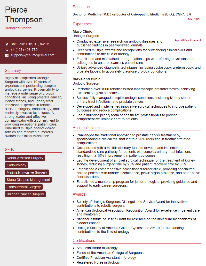
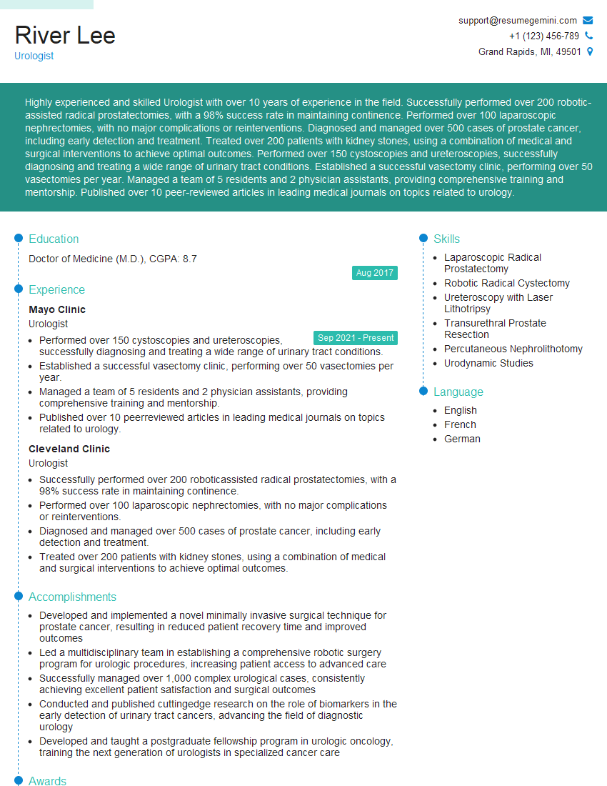
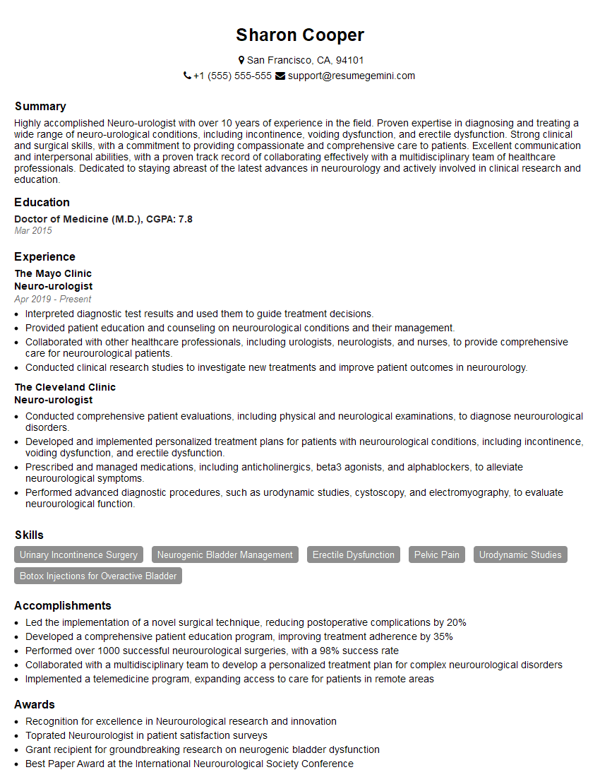
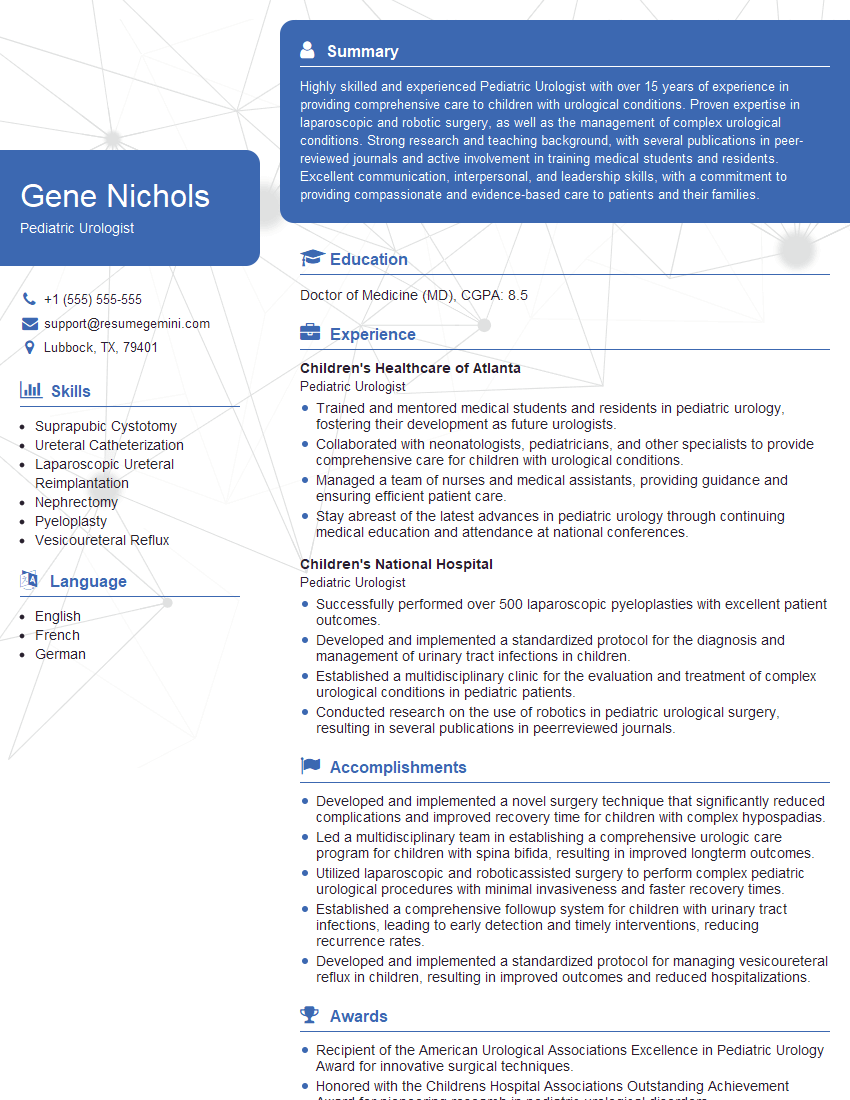
Mastering bladder surgery is crucial for career advancement in urology, opening doors to specialized fellowships, leadership roles, and increased research opportunities. To maximize your job prospects, it’s vital to present your skills and experience effectively. Creating an ATS-friendly resume is key to getting your application noticed by recruiters. We highly recommend using ResumeGemini to build a professional and impactful resume that highlights your expertise in bladder surgery. ResumeGemini provides examples of resumes tailored to bladder surgery specialists, giving you a head start in crafting your perfect application.
Explore more articles
Users Rating of Our Blogs
Share Your Experience
We value your feedback! Please rate our content and share your thoughts (optional).
What Readers Say About Our Blog
Take a look at this stunning 2-bedroom apartment perfectly situated NYC’s coveted Hudson Yards!
https://bit.ly/Lovely2BedsApartmentHudsonYards
Live Rent Free!
https://bit.ly/LiveRentFREE
Interesting Article, I liked the depth of knowledge you’ve shared.
Helpful, thanks for sharing.
Hi, I represent a social media marketing agency and liked your blog
Hi, I represent an SEO company that specialises in getting you AI citations and higher rankings on Google. I’d like to offer you a 100% free SEO audit for your website. Would you be interested?


