The right preparation can turn an interview into an opportunity to showcase your expertise. This guide to Cellular Biology interview questions is your ultimate resource, providing key insights and tips to help you ace your responses and stand out as a top candidate.
Questions Asked in Cellular Biology Interview
Q 1. Describe the process of cell signaling.
Cell signaling is the process by which cells communicate with each other and their environment. It’s a fundamental process in all living organisms, orchestrating everything from simple cell growth to complex organismal development. Think of it like a sophisticated network of messengers carrying instructions throughout a city (the organism).
The process typically involves several key steps:
- Reception: A signaling molecule (ligand) binds to a specific receptor protein on the target cell’s surface or inside the cell.
- Transduction: The binding triggers a cascade of intracellular events, often involving a series of protein modifications (like phosphorylation) that amplify the signal.
- Response: The signal ultimately leads to a specific cellular response, such as changes in gene expression, metabolism, or cell movement. This response could be anything from cell division to apoptosis (programmed cell death).
Example: Imagine insulin, a hormone that regulates blood sugar. When blood glucose is high, insulin binds to its receptors on liver cells. This triggers a cascade of intracellular signals, resulting in the uptake of glucose from the blood, lowering blood sugar levels. This is a classic example of cell signaling maintaining homeostasis.
Q 2. Explain the differences between mitosis and meiosis.
Mitosis and meiosis are both types of cell division, but they serve very different purposes. Mitosis is responsible for cell growth and repair, while meiosis produces gametes (sex cells) for sexual reproduction.
- Mitosis: This process creates two identical daughter cells from a single parent cell. Each daughter cell has the same number of chromosomes as the parent cell (diploid). Think of it as making a perfect copy – ideal for repairing damaged tissue or for growth.
- Meiosis: This process produces four genetically diverse daughter cells, each with half the number of chromosomes as the parent cell (haploid). This reduction in chromosome number is crucial for sexual reproduction, ensuring that when gametes fuse during fertilization, the resulting zygote has the correct diploid number of chromosomes. The genetic diversity arises from crossing over (exchange of genetic material between homologous chromosomes) and independent assortment (random alignment of chromosomes during metaphase I).
Key Differences Summarized:
- Number of daughter cells: Mitosis – 2; Meiosis – 4
- Chromosome number: Mitosis – diploid; Meiosis – haploid
- Genetic variation: Mitosis – none; Meiosis – significant
- Purpose: Mitosis – growth, repair; Meiosis – sexual reproduction
Q 3. What are the key components of the cell membrane and their functions?
The cell membrane, also known as the plasma membrane, is a selectively permeable barrier that encloses the cell’s contents. Its key components and their functions are:
- Phospholipids: These form a bilayer, the fundamental structure of the membrane. The hydrophobic tails face inwards, creating a barrier to water-soluble substances, while the hydrophilic heads interact with the aqueous environment inside and outside the cell.
- Cholesterol: Embedded within the phospholipid bilayer, cholesterol regulates membrane fluidity. It prevents the membrane from becoming too rigid at low temperatures or too fluid at high temperatures.
- Proteins: Membrane proteins perform a variety of functions, including:
- Transport proteins: Facilitate the movement of molecules across the membrane (e.g., ion channels, carrier proteins).
- Receptor proteins: Bind to signaling molecules, initiating intracellular signaling pathways.
- Enzymes: Catalyze reactions at the membrane surface.
- Cell adhesion molecules: Facilitate cell-cell and cell-matrix interactions.
- Carbohydrates: Often attached to lipids (glycolipids) or proteins (glycoproteins), carbohydrates play a role in cell recognition and cell signaling.
The cell membrane’s selective permeability is crucial for maintaining the cell’s internal environment, allowing essential substances to enter and waste products to exit while preventing harmful substances from entering.
Q 4. Discuss the role of the endoplasmic reticulum in protein synthesis.
The endoplasmic reticulum (ER) is a network of interconnected membranes involved in various cellular processes, notably protein synthesis. There are two main types of ER:
- Rough ER (RER): Its surface is studded with ribosomes, which are the sites of protein synthesis. Ribosomes on the RER synthesize proteins destined for secretion, membrane integration, or transport to other organelles. The RER also plays a role in protein folding and modification.
- Smooth ER (SER): Lacks ribosomes and is involved in lipid synthesis, carbohydrate metabolism, and detoxification. It’s particularly abundant in cells that specialize in these functions, such as liver cells.
Protein synthesis on the RER: Ribosomes translate mRNA into polypeptide chains. As a protein is being synthesized, if it contains a signal sequence targeting it to the ER, it will be translocated into the lumen (interior space) of the RER. Inside the RER, the protein undergoes folding and modification, such as glycosylation (addition of carbohydrate chains), before being packaged into transport vesicles for delivery to its final destination – be it the Golgi apparatus, the cell membrane, or secretion outside the cell.
Q 5. Explain the function of the Golgi apparatus.
The Golgi apparatus, also known as the Golgi complex, is a stack of flattened, membrane-bound sacs (cisternae) that acts as a processing and packaging center for proteins and lipids. Think of it as the cell’s post office, sorting and shipping packages to their correct destinations.
Its key functions include:
- Protein modification: Further processing and modification of proteins received from the ER, including glycosylation, proteolytic cleavage (cutting proteins into smaller functional units), and the addition of other molecules.
- Sorting and packaging: Sorting proteins and lipids into vesicles for transport to their correct destinations, such as the cell membrane, lysosomes, or secretion outside the cell.
- Synthesis of certain macromolecules: The Golgi apparatus also participates in the synthesis of some polysaccharides and glycolipids.
Example: Proteins synthesized in the ER, destined for secretion, are transported to the Golgi apparatus. Here, they undergo final modifications, are sorted into vesicles, and are then transported to the cell membrane for secretion outside the cell. This is how many hormones and digestive enzymes are released.
Q 6. Describe the different types of cell junctions.
Cell junctions are specialized structures that connect cells to each other or to the extracellular matrix (ECM). They provide structural support, communication pathways, and regulate the passage of molecules between cells. Different types of cell junctions exist, each with unique characteristics:
- Tight junctions: Form a watertight seal between adjacent cells, preventing the passage of molecules between them. Think of them as zippers, sealing off spaces between cells. They’re crucial in epithelial tissues lining organs and cavities.
- Adherens junctions: Connect cells via cadherin proteins, providing strong adhesion. They contribute to tissue integrity and are often found associated with the actin cytoskeleton. Imagine strong adhesive tape connecting cells.
- Desmosomes: Provide strong anchoring junctions, connecting cells via intermediate filaments. They are particularly important in tissues subject to mechanical stress, such as skin and heart muscle. These are like rivets holding the structure together.
- Gap junctions: Form channels that allow direct communication between the cytoplasm of adjacent cells. These channels allow small molecules and ions to pass freely, enabling rapid cell-to-cell signaling. They are crucial for coordinated functions in tissues like heart muscle.
- Hemidesmosomes: Anchor cells to the basal lamina (a specialized layer of the ECM). They provide a strong connection between cells and the underlying tissue.
Q 7. What are the stages of the cell cycle?
The cell cycle is an ordered series of events that leads to cell growth and division. It consists of several distinct phases:
- Interphase: This is the longest phase, where the cell grows, replicates its DNA, and prepares for division. Interphase is further divided into:
- G1 (Gap 1): Cell growth and normal metabolic activities.
- S (Synthesis): DNA replication.
- G2 (Gap 2): Further cell growth and preparation for mitosis.
- M (Mitotic) phase: This phase involves cell division, consisting of:
- Mitosis: Nuclear division (prophase, metaphase, anaphase, telophase).
- Cytokinesis: Cytoplasmic division, resulting in two separate daughter cells.
Checkpoints: The cell cycle is tightly regulated by checkpoints, ensuring that each phase is completed correctly before proceeding to the next. These checkpoints monitor DNA integrity, DNA replication, and spindle assembly, preventing errors and ensuring the fidelity of cell division. Problems at checkpoints can lead to cell cycle arrest or apoptosis.
Q 8. Explain the process of DNA replication.
DNA replication is the process by which a cell duplicates its DNA before cell division, ensuring each daughter cell receives a complete and identical copy. It’s a remarkably precise process, involving several key enzymes and steps. Imagine it like photocopying a very important document – you want an exact copy!
- Initiation: Replication begins at specific sites called origins of replication. Helicases unwind the DNA double helix, creating a replication fork.
- Unwinding and Stabilization: Single-strand binding proteins (SSBs) prevent the separated strands from re-annealing. Topoisomerases relieve the torsional stress ahead of the replication fork.
- Primer Synthesis: Primase synthesizes short RNA primers, providing a starting point for DNA polymerase.
- Elongation: DNA polymerase III adds nucleotides to the 3′ end of the RNA primer, synthesizing new DNA strands. Leading strand synthesis is continuous, while lagging strand synthesis is discontinuous, forming Okazaki fragments.
- Primer Removal and Replacement: DNA polymerase I removes RNA primers and replaces them with DNA.
- Ligation: DNA ligase joins the Okazaki fragments on the lagging strand, creating a continuous DNA molecule.
Errors during replication are rare thanks to DNA polymerase’s proofreading activity. However, errors that do occur can lead to mutations with potential consequences ranging from benign to disease-causing.
Q 9. Describe the process of transcription and translation.
Transcription and translation are the two main steps in gene expression, the process by which the information encoded in a gene is used to synthesize a functional product, usually a protein. Think of it like translating a recipe (DNA) into a delicious cake (protein).
Transcription: This process takes place in the nucleus and involves the synthesis of an RNA molecule (mRNA) from a DNA template. RNA polymerase binds to the promoter region of a gene, unwinds the DNA, and synthesizes a complementary RNA strand. This mRNA then undergoes processing (e.g., splicing) before exiting the nucleus.
Translation: This process occurs in the cytoplasm at the ribosomes. The mRNA sequence is read in codons (three-nucleotide units), each codon specifying a particular amino acid. tRNA molecules, carrying specific amino acids, recognize and bind to the corresponding codons on the mRNA. The ribosome facilitates the formation of peptide bonds between amino acids, creating a polypeptide chain that folds into a functional protein.
For example, the gene encoding the protein insulin undergoes both transcription and translation to produce the insulin hormone, which is crucial for regulating blood sugar levels.
Q 10. What are the different types of cell death?
Cell death is a fundamental process essential for development, tissue homeostasis, and the elimination of damaged or infected cells. There are two primary types of cell death: apoptosis and necrosis.
- Apoptosis (programmed cell death): This is a highly regulated and organized process characterized by specific morphological changes, such as cell shrinkage, chromatin condensation, and the formation of apoptotic bodies. Apoptosis is crucial for development (e.g., sculpting fingers and toes) and eliminating cells that are damaged or no longer needed.
- Necrosis (accidental cell death): This is an uncontrolled and often pathological form of cell death resulting from acute cellular injury, such as trauma, infection, or ischemia (lack of blood flow). It’s characterized by cell swelling, membrane rupture, and inflammation.
Other types of cell death, such as autophagy (self-eating) and pyroptosis (inflammatory cell death) also exist, with distinct mechanisms and roles in cellular processes.
Q 11. Explain the role of apoptosis in development and disease.
Apoptosis, or programmed cell death, plays a vital role in both development and disease. Its precise regulation is critical for maintaining tissue homeostasis.
In Development: Apoptosis is essential for sculpting tissues and organs. For instance, the formation of fingers and toes requires the removal of cells between the developing digits through apoptosis. Failure of apoptosis during development can lead to severe birth defects.
In Disease: Dysregulation of apoptosis can contribute to various diseases. Excessive apoptosis can result in neurodegenerative diseases such as Alzheimer’s disease. Conversely, insufficient apoptosis can lead to the development and progression of cancer, allowing damaged or abnormal cells to survive and proliferate.
Understanding the mechanisms that regulate apoptosis is crucial for developing therapeutic strategies for various diseases. For example, cancer therapies often aim to induce apoptosis in cancer cells to eliminate tumors.
Q 12. Discuss the role of mitochondria in cellular respiration.
Mitochondria are often called the ‘powerhouses’ of the cell because they are the primary site of cellular respiration, the process of generating ATP (adenosine triphosphate), the main energy currency of the cell. This process involves several key steps:
- Glycolysis: Glucose is broken down in the cytoplasm, yielding pyruvate.
- Pyruvate Oxidation: Pyruvate enters the mitochondria and is converted to acetyl-CoA.
- Krebs Cycle (Citric Acid Cycle): Acetyl-CoA enters the Krebs cycle, a series of reactions that produce NADH, FADH2, and ATP.
- Oxidative Phosphorylation: This is the final and most energy-yielding stage. Electrons from NADH and FADH2 are passed along the electron transport chain, embedded in the inner mitochondrial membrane. This process generates a proton gradient, which drives ATP synthesis by ATP synthase.
Mitochondria also play crucial roles in other cellular processes, including calcium homeostasis, apoptosis, and the synthesis of certain metabolites. Mitochondrial dysfunction is implicated in a wide range of diseases, including neurodegenerative disorders and metabolic syndromes.
Q 13. What are the different types of microscopy used in cell biology?
Various microscopy techniques are used in cell biology to visualize cells and their components at different magnifications and resolutions. The choice of microscopy depends on the specific research question and the structures of interest.
- Light Microscopy: This is the most common type, using visible light to illuminate the sample. Different types of light microscopy exist, including bright-field, phase-contrast, fluorescence, and confocal microscopy.
- Electron Microscopy: This technique uses a beam of electrons instead of light, providing much higher resolution. Transmission electron microscopy (TEM) allows visualization of internal cellular structures, while scanning electron microscopy (SEM) provides high-resolution images of cell surfaces.
- Super-resolution Microscopy: Techniques such as PALM and STORM overcome the diffraction limit of light microscopy, enabling visualization of structures smaller than the wavelength of light.
Each type of microscopy has its strengths and limitations, making it crucial to select the appropriate technique for the research objective.
Q 14. Explain the principles of flow cytometry.
Flow cytometry is a powerful technique used to analyze the physical and chemical characteristics of individual cells within a heterogeneous population. It’s like sorting candies based on their color and size – each candy representing a cell.
Cells are suspended in a fluid and passed through a laser beam one at a time. The scattered light and emitted fluorescence are measured by detectors, providing information about cell size, granularity, and the presence of specific surface markers or intracellular molecules. This data is then used to sort and analyze cells based on their properties. For example, flow cytometry can be used to identify and isolate specific immune cells, such as T cells or B cells, based on the expression of specific surface markers.
Flow cytometry has wide applications in various fields, including immunology, hematology, cancer biology, and microbiology. It’s particularly useful for identifying and isolating rare cell populations, studying cell cycle progression, and analyzing the expression of various proteins.
Q 15. Describe various cell culture techniques.
Cell culture is the process of growing cells outside of their natural environment, typically in a controlled laboratory setting. This allows researchers to study cellular processes, test drug efficacy, and engineer tissues. Various techniques exist, tailored to different cell types and experimental goals.
- Primary Cell Culture: Cells are isolated directly from a tissue and cultured. These cells have a limited lifespan and tend to retain characteristics of their tissue of origin. Think of it like harvesting apples directly from a tree – you get the freshest, most authentic product, but it won’t last forever.
- Cell Lines: These are immortalized cells that can divide indefinitely in culture. They are often derived from primary cultures or tumors and are easier to maintain than primary cells. A cell line is more like a cultivated apple orchard—the apples are less ‘natural’ but you can harvest continuously.
- Organotypic Cultures: These cultures attempt to mimic the three-dimensional structure and interactions of cells within a tissue or organ. This can be achieved using techniques like scaffold-based culture or spheroid formation. This represents the next level – creating a small ‘apple tree’ in a controlled environment.
- Suspension Culture: Cells are grown in suspension, without attachment to a surface. This is common for blood cells and some cancer cells. Think of growing apples in a large water tank, supported solely by the liquid.
- Adherent Culture: Cells require attachment to a surface (typically a culture dish treated with materials like collagen or fibronectin) to grow and proliferate. This is the most common method for many cell types; like how most apple trees need to be planted in soil.
Career Expert Tips:
- Ace those interviews! Prepare effectively by reviewing the Top 50 Most Common Interview Questions on ResumeGemini.
- Navigate your job search with confidence! Explore a wide range of Career Tips on ResumeGemini. Learn about common challenges and recommendations to overcome them.
- Craft the perfect resume! Master the Art of Resume Writing with ResumeGemini’s guide. Showcase your unique qualifications and achievements effectively.
- Don’t miss out on holiday savings! Build your dream resume with ResumeGemini’s ATS optimized templates.
Q 16. How do you design and execute a cell-based assay?
Designing and executing a cell-based assay involves a series of carefully considered steps, from hypothesis formation to data analysis. A crucial aspect is ensuring reproducibility and minimizing experimental error.
- Define the objective: What biological process or effect are you trying to measure?
- Choose the appropriate cell type: Select a cell line that is relevant to your research question and exhibits suitable characteristics (e.g., expression of target proteins, responsiveness to treatments).
- Assay design: Determine the specific assay method (e.g., cell viability assay, proliferation assay, ELISA). Consider controls (positive and negative), sample size, and potential confounding factors.
- Cell culture: Ensure consistent and healthy cell cultures by maintaining appropriate conditions (temperature, humidity, CO2). Perform necessary quality checks (e.g., cell morphology, viability).
- Treatment and incubation: Apply treatments (e.g., drugs, siRNA, growth factors) as per your experimental design, ensuring consistent application and timing.
- Data acquisition and analysis: Collect data according to the assay method. Perform appropriate statistical analyses to determine significance and draw conclusions.
- Validation and reproducibility: Repeat experiments independently to ensure reproducibility and reliability of results.
For example, if investigating the effect of a new drug on cell proliferation, a viable assay would include a cell counting kit-8 (CCK-8) assay. You would compare treated cells against untreated control groups, ensuring statistically significant differences support your conclusions.
Q 17. Explain your understanding of CRISPR-Cas9 gene editing.
CRISPR-Cas9 is a revolutionary gene-editing technology derived from a bacterial defense system. It allows researchers to precisely target and modify specific DNA sequences within a genome.
The system comprises two key components:
- Cas9: An enzyme that acts as molecular scissors, cutting DNA at a specific location.
- Guide RNA (gRNA): A short RNA molecule that directs Cas9 to the target DNA sequence through base pairing. The gRNA sequence must be carefully designed to be specific to the desired target.
The process involves designing a gRNA targeting a specific gene, delivering both the gRNA and Cas9 into the cell (often using viral vectors or transfection methods), and allowing Cas9 to cleave the DNA at the targeted site. This double-strand break can then be repaired by the cell through two primary mechanisms:
- Non-homologous end joining (NHEJ): An error-prone repair pathway often leading to insertions or deletions, causing gene disruption (gene knockout).
- Homology-directed repair (HDR): A more precise repair pathway that uses a provided DNA template to repair the break, allowing for precise gene editing (gene knockin or correction).
CRISPR-Cas9 has broad applications in biomedical research, including disease modeling, gene therapy, and drug discovery. However, off-target effects (unintended edits at other genomic locations) remain a challenge that requires careful consideration and optimization.
Q 18. Discuss the ethical considerations of stem cell research.
Stem cell research presents numerous ethical considerations, primarily revolving around the source of the stem cells and their potential applications.
- Embryonic Stem Cells: Derived from human embryos, their use raises concerns about the destruction of potential human life. The debate centers around the moral status of the embryo and the balance between potential medical benefits and ethical objections.
- Induced Pluripotent Stem Cells (iPSCs): These are adult cells reprogrammed to an embryonic-like pluripotent state, bypassing the ethical concerns associated with embryonic stem cells. However, there are still questions surrounding the safety and efficacy of reprogramming techniques and potential long-term effects.
- Somatic Stem Cells: These adult stem cells are found in various tissues and have limited pluripotency, reducing ethical concerns compared to embryonic stem cells. However, their limited differentiation potential may restrict their therapeutic applications.
- Informed Consent: Whenever human cells are used in research, informed consent is absolutely crucial, ensuring donors understand the procedures and implications of their contribution. Maintaining donor anonymity and data privacy are also paramount.
- Therapeutic Applications: The potential for stem cells to treat a wide range of diseases raises ethical considerations regarding access, cost, and equitable distribution of these therapies.
Regulatory oversight, ongoing dialogue between scientists, ethicists, and the public, and the development of strict ethical guidelines are crucial for responsible and beneficial stem cell research.
Q 19. Describe the methods for isolating and culturing specific cell types.
Isolating and culturing specific cell types often requires a combination of techniques designed to separate the target cells from other cell types within a tissue sample.
- Enzymatic Digestion: Tissues are treated with enzymes (e.g., collagenase, trypsin) that break down the extracellular matrix, releasing individual cells.
- Mechanical Dissociation: Physical methods (e.g., pipetting, trituration) can be used to separate cells, especially in tissues with less dense extracellular matrix.
- Density Gradient Centrifugation: This technique uses density gradients to separate cells based on their size and density. For example, isolating lymphocytes from peripheral blood uses Ficoll gradient.
- Fluorescence-Activated Cell Sorting (FACS): This powerful technique uses fluorescently labeled antibodies to identify and sort specific cell types based on surface markers. This is crucial for isolating rare cell populations.
- Magnetic-Activated Cell Sorting (MACS): Similar to FACS, but uses magnetic beads coated with antibodies to isolate cells. This method is less expensive than FACS but potentially has lower resolution.
- Micromanipulation: Individual cells can be selected and isolated using specialized microscopes and tools. This method is typically used for rare cells or when high purity is required.
Once isolated, cells are cultured using appropriate media and conditions, often supplemented with growth factors or other molecules necessary for their survival and proliferation. The choice of culture techniques (adherent vs. suspension) depends on the cell type and its growth requirements.
Q 20. How would you troubleshoot a failed cell culture experiment?
Troubleshooting a failed cell culture experiment involves systematic investigation of potential causes, starting with the most common issues.
- Contamination: Check for bacterial, fungal, or mycoplasma contamination. This can be done microscopically and with appropriate assays. Contamination is often characterized by turbidity in the media, unusual cell morphology, or rapid cell death.
- Incubation Conditions: Verify correct temperature, CO2 levels, and humidity are maintained in the incubator. Fluctuations can severely impact cell growth.
- Media and Reagents: Inspect the media for signs of degradation or contamination. Check the expiry dates of all reagents. Ensure proper storage and handling procedures.
- Cell Health and Viability: Examine the cells microscopically for signs of stress (e.g., rounding, detachment). Assess cell viability using appropriate assays (e.g., trypan blue exclusion).
- Subculturing Technique: If subculturing was involved, verify the correct enzymatic treatment, cell seeding density, and passage number were used. Improper subculturing can lead to cell death or poor growth.
- Experimental Protocol: Carefully review the experimental procedure for any errors or omissions. Consider alternative protocols if necessary.
Systematic troubleshooting, meticulous record-keeping, and use of control groups are crucial in identifying the root cause of the failure and preventing recurrence.
Q 21. Explain your experience with various types of cellular imaging.
Cellular imaging is a fundamental tool in cellular biology, providing visual information about cell structure, function, and behavior. Various techniques provide different levels of detail and information.
- Brightfield Microscopy: A basic technique providing visualization of cell morphology using transmitted light. Suitable for observing cell shape and size but lacks detailed intracellular information.
- Fluorescence Microscopy: Uses fluorescent probes (e.g., fluorescent proteins, antibodies) to visualize specific molecules or structures within cells. Confocal microscopy enhances resolution by eliminating out-of-focus light.
- Electron Microscopy: Provides high-resolution images, capable of visualizing cellular ultrastructure (e.g., organelles, membranes). Transmission electron microscopy (TEM) is used for thin sections, while scanning electron microscopy (SEM) is used for surface imaging.
- Live-Cell Imaging: Observing cells over time allows the dynamics of cellular processes to be studied (e.g., cell migration, division, intracellular trafficking). Requires specialized incubators and imaging systems.
- Super-resolution Microscopy: Techniques like PALM and STORM overcome the diffraction limit of light, achieving resolutions beyond the capabilities of conventional light microscopy, enabling visualization of nanoscale structures.
The choice of imaging technique depends on the specific research question, the resolution required, and the nature of the cellular structures or processes under investigation. Proper sample preparation and image analysis are critical for obtaining meaningful results.
Q 22. How would you interpret Western blot results?
Interpreting Western blot results involves analyzing protein expression levels. A Western blot is like a detective’s tool to identify a specific protein within a complex mixture of proteins extracted from cells or tissues. First, you’ll see the bands on the blot – darker bands indicate higher protein levels, while faint or absent bands mean low or no protein expression. The size of the protein is determined by its migration distance relative to a molecular weight marker ladder also run on the gel.
Key steps to interpretation:
- Check the ladder: This helps determine the molecular weight of your target protein.
- Identify target protein band: Look for a band at the expected molecular weight.
- Compare across samples: Quantify band intensity using densitometry software (ImageJ is a common choice) to compare protein expression levels across different samples (e.g., treated vs. untreated cells, different cell lines). A significant difference in band intensity suggests a change in protein expression.
- Control samples are essential: These ensure the experiment worked correctly. A loading control (e.g., β-actin) verifies equal protein loading in each lane. A positive control confirms the antibody’s specificity.
Example: If you’re investigating a drug’s effect on a specific protein, and you see a significant decrease in band intensity in the drug-treated samples compared to the control, that suggests the drug is effectively reducing the expression of that protein.
Q 23. What is your experience with data analysis in the context of cellular biology?
My experience with data analysis in cellular biology is extensive, encompassing various techniques and software. I’m proficient in analyzing data from a wide range of experiments including Western blots, flow cytometry, qPCR, and microscopy. My workflow typically includes data cleaning, normalization, statistical analysis, and visualization.
Specifically, I’ve used statistical software like GraphPad Prism and R for hypothesis testing (t-tests, ANOVA, etc.), curve fitting, and correlation analysis. I’m familiar with various bioinformatics tools for analyzing genomic and proteomic data. For example, in one project involving RNA sequencing, I used tools like DESeq2 in R to identify differentially expressed genes after treatment with a novel compound.
I find data visualization crucial for communicating results clearly. I use various software like GraphPad Prism, R (ggplot2 package), and other tools to generate publication-quality figures effectively illustrating my findings. I believe a solid understanding of statistical methods is crucial to avoid misinterpretations and draw accurate conclusions from experimental data.
Q 24. Describe a challenging research project you worked on and how you overcame obstacles.
One challenging project involved investigating the role of a specific microRNA in cancer cell metastasis. Initially, our attempts to knock down the microRNA using traditional siRNA methods proved largely ineffective. We consistently observed very low knockdown efficiency despite several optimization attempts.
The obstacle was the inherent instability of the siRNA and off-target effects. To overcome this, we explored alternative approaches. We switched to using CRISPR-Cas9 technology for gene editing, which provided a more efficient and specific method to target the microRNA. We designed specific guide RNAs targeting the microRNA gene and transfected them into cancer cell lines. We carefully monitored the transfection efficiency and performed rigorous validation experiments to ensure specificity.
This involved multiple rounds of optimization, including adjusting the concentration of guide RNA, CRISPR components, and transfection methods. We carefully monitored off-target effects and employed several methods to validate the knockdown. Ultimately, this yielded successful and reproducible knockdown, allowing us to proceed with our experiments and determine the specific role of the microRNA in the metastatic process. This experience emphasized the importance of adopting a flexible approach, exploring alternative techniques, and meticulous validation in research.
Q 25. Explain your understanding of different types of cell lines and their characteristics.
Cell lines are immortalized cells grown in the lab, offering a consistent experimental model. However, they have limitations since they don’t perfectly reflect the complexity of in vivo systems. There are several types:
- Primary cell lines: Derived directly from tissues. They have a limited lifespan and are more representative of in vivo cells, though more challenging to work with.
- Immortalized cell lines: Genetically modified to have unlimited replication potential. HeLa cells are a classic example. They are easy to culture but can display altered phenotypes compared to their primary counterparts.
- Cancer cell lines: Derived from tumors. These reflect some characteristics of cancer cells, offering valuable models for cancer research. They can vary significantly in their genetic and phenotypic properties.
- Stem cell lines: Can differentiate into various cell types. Embryonic stem cells (ESCs) and induced pluripotent stem cells (iPSCs) are examples.
Characteristics vary greatly: Each cell line exhibits unique properties like growth rate, morphology, and genetic makeup. Understanding these characteristics is crucial for selecting the appropriate cell line for an experiment and interpreting the results. For example, a fast-growing cell line might not be suitable for studying the long-term effects of a drug, while a specific cancer cell line might be ideal for testing an anti-cancer compound.
Q 26. How would you design an experiment to investigate the effects of a drug on cell proliferation?
To investigate a drug’s effect on cell proliferation, I’d design an experiment incorporating several key controls and techniques:
- Cell culture: Select an appropriate cell line relevant to the drug’s target. Maintain consistent cell culture conditions to ensure reproducibility.
- Drug treatment: Treat cells with various concentrations of the drug, including a control group receiving vehicle (e.g., DMSO) only. A dose-response curve should be established to determine the optimal concentration range.
- Proliferation assays: Several assays can measure cell proliferation. Examples include:
- MTT assay: Measures metabolic activity, reflecting cell viability and proliferation.
- Cell counting: Direct counting of cells using a hemocytometer or automated cell counter at various time points.
- Colony formation assay: Assesses the ability of cells to form colonies, indicating long-term proliferation.
- EdU incorporation assay: Measures DNA synthesis during S-phase of the cell cycle.
- Data analysis: Analyze the results using appropriate statistical tests (e.g., t-test, ANOVA) to compare cell proliferation between drug-treated and control groups. Create graphs to visually present the data (dose-response curves, bar graphs).
- Validation: Incorporate additional assays (e.g., flow cytometry to assess cell cycle progression or apoptosis) to confirm the results from the proliferation assay.
The overall experimental design will ensure that the observed changes in cell proliferation are directly attributable to the drug and not other factors. For example, controls for cell seeding density and media consistency would be crucial.
Q 27. Discuss the impact of epigenetic modifications on gene expression.
Epigenetic modifications are heritable changes in gene expression that do not involve alterations to the underlying DNA sequence. They act as a layer of regulation on top of the genetic code, influencing which genes are turned ‘on’ or ‘off’. These modifications play a crucial role in development, cell differentiation, and disease.
Key types of epigenetic modifications:
- DNA methylation: The addition of a methyl group (CH3) to cytosine bases in DNA. Usually associated with gene silencing.
- Histone modification: Chemical modifications (e.g., acetylation, methylation, phosphorylation) to histone proteins around which DNA is wrapped. These modifications can alter chromatin structure, influencing gene accessibility.
- Non-coding RNAs (ncRNAs): These RNA molecules do not code for proteins but regulate gene expression through various mechanisms, like targeting mRNA for degradation or inhibiting translation.
Impact on gene expression:
- Gene silencing: DNA methylation and certain histone modifications (e.g., histone deacetylation) can condense chromatin, making genes inaccessible to the transcriptional machinery and resulting in gene silencing.
- Gene activation: Histone acetylation and other modifications can loosen chromatin structure, facilitating gene transcription and activation.
Real-world implications: Epigenetic alterations are implicated in many diseases, including cancer. Cancer cells often exhibit aberrant DNA methylation patterns and histone modifications that contribute to uncontrolled cell growth. Understanding these epigenetic changes is crucial for developing targeted therapies.
Key Topics to Learn for Your Cellular Biology Interview
- Cell Structure & Function: Understand the intricacies of organelles, their roles, and interactions. Consider practical applications in drug delivery and disease mechanisms.
- Membrane Biology: Grasp concepts of membrane transport (passive and active), signal transduction pathways, and their relevance to cellular communication and disease states. Explore techniques like fluorescence microscopy and patch clamping.
- Cell Signaling: Master various signaling pathways (e.g., G-protein coupled receptors, receptor tyrosine kinases) and their roles in regulating cellular processes. Think about the implications for cancer research and therapeutic development.
- Cell Cycle & Cell Division: Understand the phases of the cell cycle, checkpoints, and regulation. Consider practical applications in cancer biology and drug development targeting cell division.
- Cellular Metabolism: Explore glycolysis, oxidative phosphorylation, and other metabolic pathways. Think about the applications in metabolic diseases and the development of metabolic therapies.
- Gene Expression & Regulation: Understand transcription, translation, and post-translational modifications. Consider the implications for genetic engineering and gene therapy.
- Cellular Respiration: Understand the process of cellular respiration and its importance in energy production. Consider the practical implications for understanding mitochondrial diseases.
- Apoptosis and Necrosis: Distinguish between programmed cell death (apoptosis) and accidental cell death (necrosis) and their implications for disease and development.
- Modern Techniques in Cellular Biology: Be familiar with common techniques like microscopy (confocal, electron), flow cytometry, PCR, and Western blotting. Prepare to discuss their principles and applications.
Next Steps
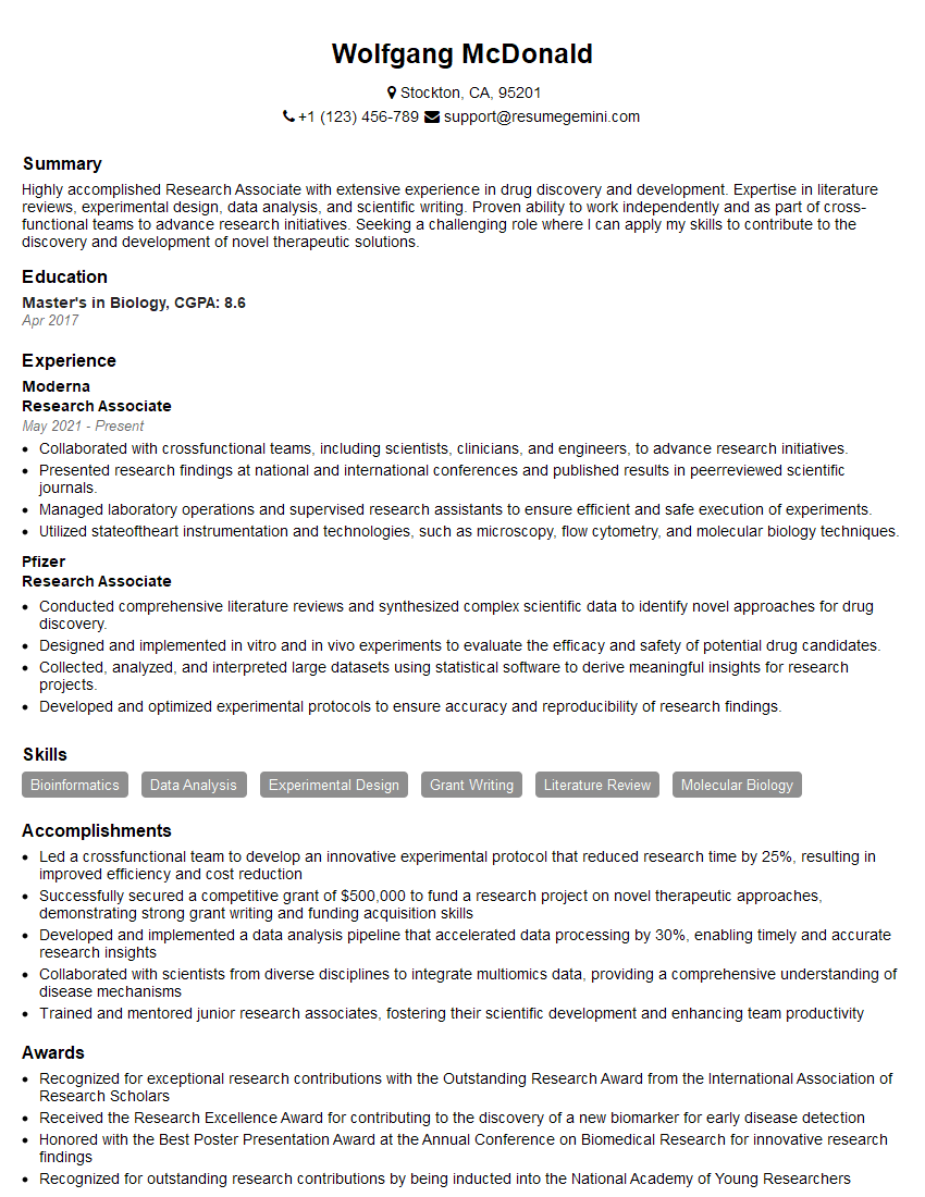
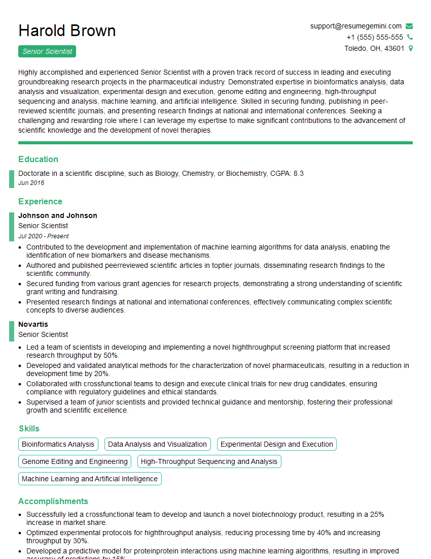
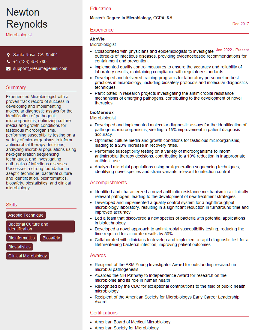
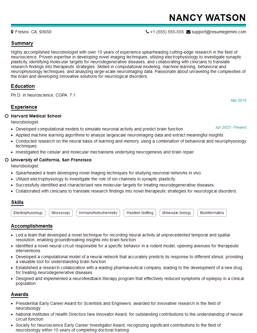
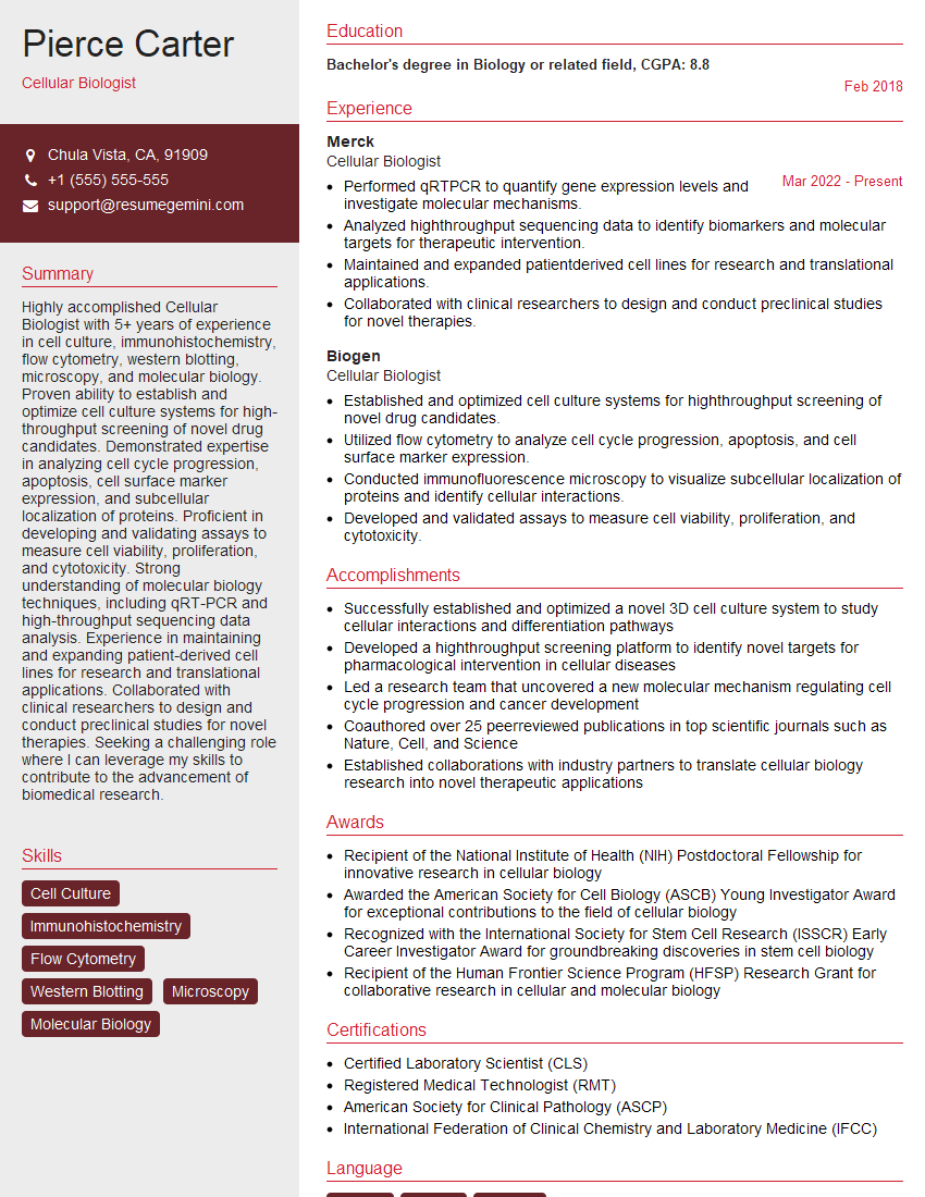
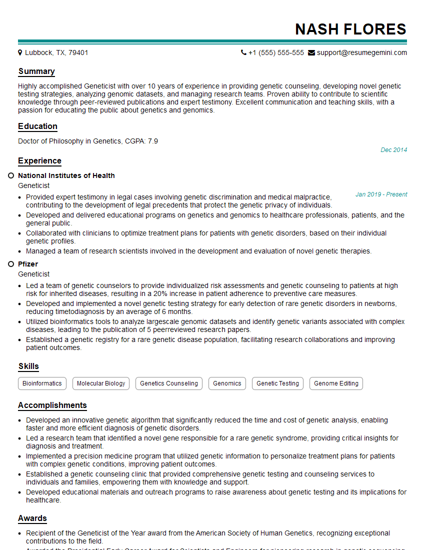
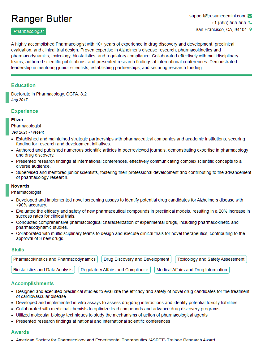
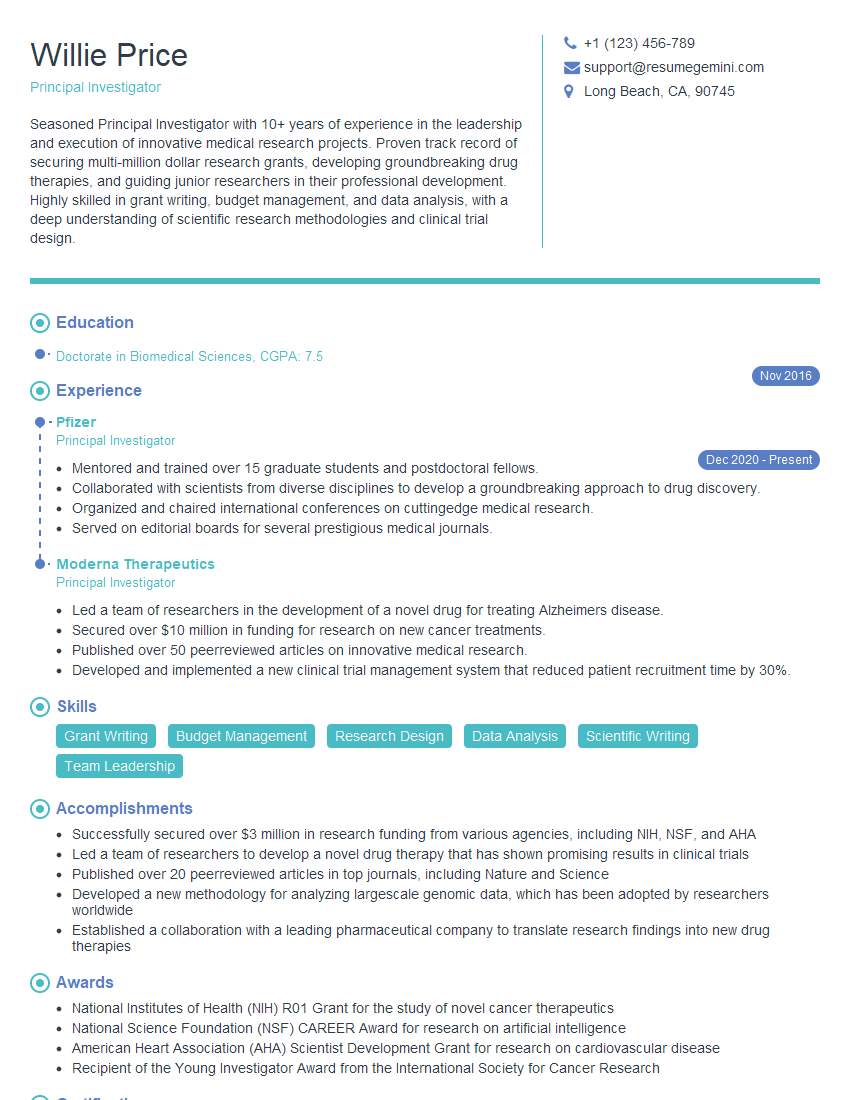
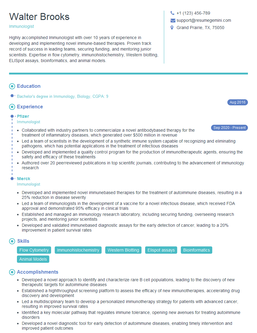
Mastering Cellular Biology opens doors to exciting career paths in research, pharmaceuticals, biotechnology, and academia. A strong understanding of these core concepts significantly enhances your interview performance and career prospects. To make a strong impression, it’s vital to have an ATS-friendly resume that showcases your skills and experience effectively. ResumeGemini is a trusted resource to help you craft a professional and impactful resume tailored to your specific career goals. We provide examples of resumes specifically designed for Cellular Biology professionals to help you get started.
Explore more articles
Users Rating of Our Blogs
Share Your Experience
We value your feedback! Please rate our content and share your thoughts (optional).
What Readers Say About Our Blog
hello,
Our consultant firm based in the USA and our client are interested in your products.
Could you provide your company brochure and respond from your official email id (if different from the current in use), so i can send you the client’s requirement.
Payment before production.
I await your answer.
Regards,
MrSmith
These apartments are so amazing, posting them online would break the algorithm.
https://bit.ly/Lovely2BedsApartmentHudsonYards
Reach out at [email protected] and let’s get started!
Take a look at this stunning 2-bedroom apartment perfectly situated NYC’s coveted Hudson Yards!
https://bit.ly/Lovely2BedsApartmentHudsonYards
Live Rent Free!
https://bit.ly/LiveRentFREE
Interesting Article, I liked the depth of knowledge you’ve shared.
Helpful, thanks for sharing.
Hi, I represent a social media marketing agency and liked your blog
Hi, I represent an SEO company that specialises in getting you AI citations and higher rankings on Google. I’d like to offer you a 100% free SEO audit for your website. Would you be interested?
