Interviews are opportunities to demonstrate your expertise, and this guide is here to help you shine. Explore the essential Urologic Surgery interview questions that employers frequently ask, paired with strategies for crafting responses that set you apart from the competition.
Questions Asked in Urologic Surgery Interview
Q 1. Describe your experience with laparoscopic pyeloplasty.
Laparoscopic pyeloplasty is a minimally invasive surgical technique used to correct ureteropelvic junction (UPJ) obstruction, a condition where the connection between the kidney and ureter is narrowed, hindering urine flow. My experience encompasses a significant number of these procedures, utilizing both transperitoneal and retroperitoneal approaches. The transperitoneal approach involves accessing the kidney through the abdominal cavity, while the retroperitoneal approach avoids entering the abdominal cavity, minimizing potential complications. I’ve found the laparoscopic approach offers numerous advantages over traditional open surgery, including smaller incisions, reduced pain, shorter hospital stays, and faster recovery times. A typical procedure involves dissecting the UPJ, identifying the stricture, and performing a pyeloplasty, which involves reconstructing the junction to restore normal urine flow. Post-operative management includes careful monitoring of urine output, pain management, and early mobilization. I’ve successfully treated a wide range of UPJ obstruction severities, from mild to severe, adapting my surgical technique as needed. For example, in cases with complex anatomical variations or significant scarring, I’ve utilized advanced techniques like robotic-assisted laparoscopic pyeloplasty for enhanced precision and visualization.
Q 2. Explain the different types of urinary incontinence and their management.
Urinary incontinence is the involuntary leakage of urine. It’s classified into several types, primarily:
- Stress incontinence: Urine leaks during physical activity or exertion, such as coughing, sneezing, or lifting. This is often caused by weakened pelvic floor muscles. Think of it like a leaky faucet – pressure causes leakage.
- Urge incontinence: A sudden, strong urge to urinate followed by involuntary leakage. This is frequently associated with overactive bladder. Imagine a fire alarm going off unexpectedly – you have to respond quickly and sometimes it’s too late to control.
- Overflow incontinence: Urine leaks due to a bladder that’s always full and unable to empty completely. This often results from bladder outlet obstruction or nerve damage. It’s similar to a constantly overflowing bathtub.
- Mixed incontinence: A combination of stress and urge incontinence.
- Functional incontinence: Leakage due to physical limitations or cognitive impairments that prevent a person from reaching the toilet in time. This isn’t a bladder problem, but a mobility or cognitive issue.
Q 3. How do you manage a patient with acute urinary retention?
Acute urinary retention (AUR) is a sudden inability to urinate. It’s a urologic emergency requiring immediate intervention. Management begins with assessing the patient’s condition – vital signs, abdominal examination for distension, and obtaining a detailed history. The first step is usually catheterization to relieve the bladder distension. A Foley catheter is typically placed to drain the urine. Once the bladder is emptied, the underlying cause needs to be investigated. This may involve blood tests, urine analysis, ultrasound, or other imaging studies to identify potential causes such as benign prostatic hyperplasia (BPH), prostate cancer, urinary tract infection (UTI), neurogenic bladder, or medication side effects. Treatment targets the root cause. For BPH, options range from medication to minimally invasive procedures like transurethral resection of the prostate (TURP). UTIs require antibiotics. Neurogenic bladder might need medication or intermittent self-catheterization. Close monitoring post-catheterization is vital to ensure adequate urine output and to identify and treat any complications.
Q 4. What are the indications for radical prostatectomy?
Radical prostatectomy is the surgical removal of the prostate gland and surrounding tissues. Indications for this procedure are primarily focused on prostate cancer. The decision for surgery is made based on several factors including:
- Stage of cancer: Early-stage, localized prostate cancer is a key indication.
- Grade of cancer: Higher-grade cancers (more aggressive) are more likely to be treated surgically.
- Patient’s overall health and life expectancy: Surgery carries risks, so the patient’s general health is a critical consideration.
- Patient preference: The patient’s wishes and understanding of the risks and benefits are paramount.
- Failure of other treatments: If other treatments like radiation therapy haven’t been successful or are unsuitable, radical prostatectomy might be considered.
Q 5. Discuss the surgical approach and potential complications of a nephrectomy.
Nephrectomy is the surgical removal of a kidney. The surgical approach depends on several factors, including the reason for the nephrectomy (e.g., cancer, trauma, kidney disease), the size and location of the kidney, and the surgeon’s expertise. Approaches include:
- Open nephrectomy: A larger incision is made to access and remove the kidney. This is typically used for large tumors or complex cases.
- Laparoscopic nephrectomy: Several small incisions are made, and specialized instruments are used to remove the kidney. This minimally invasive technique offers advantages such as smaller incisions, less pain, and faster recovery.
- Robotic-assisted laparoscopic nephrectomy: Similar to laparoscopic nephrectomy, but with robotic assistance for enhanced precision and dexterity.
Q 6. Explain your understanding of different types of kidney stones and their treatment.
Kidney stones, also known as nephrolithiasis, are hard deposits that form in the kidneys. They are broadly classified by their composition:
- Calcium stones: The most common type, often composed of calcium oxalate or calcium phosphate.
- Struvite stones: Associated with urinary tract infections caused by certain bacteria, often forming large staghorn calculi that fill the renal pelvis.
- Uric acid stones: Occur in individuals with high uric acid levels, often related to diet or medical conditions like gout.
- Cystine stones: A rare type associated with a genetic disorder called cystinuria.
Q 7. Describe the management of benign prostatic hyperplasia (BPH).
Benign prostatic hyperplasia (BPH) is a non-cancerous enlargement of the prostate gland, common in older men. Management aims to relieve urinary symptoms and prevent complications. Options range from watchful waiting (monitoring symptoms without treatment), medication, and minimally invasive or surgical procedures. Medication options include alpha-blockers (relax the prostate muscles) and 5-alpha-reductase inhibitors (shrink the prostate). Minimally invasive procedures include transurethral microwave thermotherapy (TUMT), transurethral needle ablation (TUNA), and laser therapies. Surgical options, such as transurethral resection of the prostate (TURP) or open prostatectomy, are reserved for men with significant symptoms unresponsive to medical management or minimally invasive procedures. The choice of treatment depends on the severity of symptoms, the patient’s overall health, and his preferences. Regular follow-up monitoring is crucial to assess treatment effectiveness and address any emerging issues.
Q 8. What are the diagnostic tools used in evaluating erectile dysfunction?
Evaluating erectile dysfunction (ED) requires a multi-faceted approach, combining the patient’s history with a thorough physical examination and various diagnostic tests. The goal is to identify the underlying cause, whether it’s vascular, neurological, hormonal, or psychological.
Medical History: A detailed history including age, overall health, medications (e.g., blood pressure medications, antidepressants), past medical conditions (e.g., diabetes, hypertension), and lifestyle factors (e.g., smoking, alcohol consumption, exercise) is crucial. We also explore the patient’s sexual history, including the onset, frequency, and severity of ED, as well as any associated symptoms (e.g., decreased libido, pain during intercourse).
Physical Examination: A physical exam assesses general health and focuses on the vascular and neurological systems. This includes checking for peripheral artery disease (PAD), which can impact penile blood flow. We also assess testicular size and consistency, as hormonal issues can contribute to ED.
Nocturnal Penile Tumescence (NPT) Testing: This test measures penile rigidity during sleep, helping determine if erectile function is possible. This can be helpful in differentiating between psychological and organic causes of ED.
Doppler Ultrasound: This assesses blood flow into the penis, helping identify vascular issues. We can visualize the arteries and veins to determine if there are blockages or abnormalities.
Biothesiometry: This measures penile sensory function, useful in identifying neurological causes.
Hormone Testing: Blood tests measuring testosterone, prolactin, and other hormones can help identify hormonal imbalances.
Psychosocial Evaluation: Addressing psychological factors like stress, anxiety, and relationship problems is often crucial, and may involve referral to a therapist or counselor.
For example, a patient presenting with ED and a history of diabetes may undergo Doppler ultrasound to check for vascular compromise. A patient with sudden-onset ED might undergo NPT testing to rule out a neurological cause. A comprehensive evaluation ensures we address both the physical and psychological aspects of ED.
Q 9. Discuss the role of robotic surgery in urologic oncology.
Robotic surgery has revolutionized urologic oncology, offering several advantages over traditional open surgery and laparoscopic techniques. The enhanced dexterity, precision, and 3D visualization provided by the robotic system translate to smaller incisions, less blood loss, reduced pain, shorter hospital stays, faster recovery times, and often improved cosmetic outcomes.
Radical Prostatectomy: Robotic-assisted radical prostatectomy (RARP) is now a widely accepted standard of care for prostate cancer. The precision of robotic instruments allows for meticulous dissection of the neurovascular bundles, which are responsible for erectile function, potentially preserving sexual function. This leads to improved continence and potency rates compared to open surgery in experienced hands.
Partial Nephrectomy: Robotic partial nephrectomy is becoming increasingly common for the treatment of kidney cancer. The surgeon can precisely remove the tumor while preserving as much healthy kidney tissue as possible. This minimally invasive approach minimizes blood loss and allows for faster recovery compared to traditional open surgery.
Radical Cystectomy: Robotic radical cystectomy, the removal of the bladder, is increasingly utilized in the management of bladder cancer. The improved visualization and dexterity enable precise dissection of complex pelvic anatomy, making it feasible for larger and more complex tumors, and can help preserve other organs better.
Nephroureterectomy: Robotic nephroureterectomy, the removal of the kidney and ureter, is used for tumors in the ureter or kidney, and offers the same benefits of minimally invasive surgery as other robotic procedures.
However, it’s important to note that robotic surgery requires specialized training and equipment, and its use should be tailored to the patient’s individual needs and the surgeon’s expertise. Not all urologic oncology patients are candidates for robotic surgery.
Q 10. How do you assess and manage a patient with bladder cancer?
Managing bladder cancer involves a systematic approach focusing on accurate diagnosis, staging, and treatment tailored to the individual patient. The initial assessment is crucial in determining the extent of the disease and selecting the appropriate treatment strategy.
Assessment: This begins with a thorough history, including urinary symptoms (e.g., hematuria, frequency, urgency), and a physical examination.
Cystoscopy: A cystoscopy, where a thin, flexible tube with a camera is inserted into the bladder, allows direct visualization of the bladder lining. It aids in identifying and characterizing bladder tumors, often obtaining biopsies during the procedure for further pathological analysis.
Imaging: Imaging studies such as CT scans and MRI scans play a crucial role in staging the cancer, determining the extent of local invasion and the presence of distant metastases.
Biopsy: Tissue samples are obtained via cystoscopy or transurethral resection of bladder tumor (TURBT) and sent for pathological analysis, which determines the grade and stage of the cancer. The grade refers to how aggressive the cancer cells look under a microscope and the stage describes how far the cancer has spread.
Treatment: Treatment options depend heavily on the stage and grade of the cancer. Early-stage, non-muscle invasive bladder cancers (NMIBC) might be treated with transurethral resection of the bladder tumor (TURBT), followed by intravesical chemotherapy or immunotherapy (instilling medication directly into the bladder). Muscle-invasive bladder cancers (MIBC) usually require radical cystectomy (surgical removal of the bladder and surrounding lymph nodes), often followed by adjuvant chemotherapy or radiation therapy. Advanced bladder cancer, particularly with distant metastases, may require systemic chemotherapy.
For instance, a patient with a small, low-grade NMIBC discovered during a cystoscopy might be treated with TURBT and intravesical BCG (Bacillus Calmette-Guérin) immunotherapy. Conversely, a patient with a high-grade MIBC and lymph node involvement would require a radical cystectomy and potentially chemotherapy.
Q 11. Explain your approach to managing a patient with ureteral stricture.
Managing ureteral strictures, or narrowings of the ureter, requires a careful evaluation and a tailored approach depending on the severity, location, and cause of the stricture. The goal is to restore normal urine flow and prevent kidney damage from obstruction.
Assessment: Diagnosis typically involves a detailed history and physical examination, along with imaging studies such as intravenous pyelography (IVP), CT urography, or MRI urography to visualize the urinary tract and identify the location and extent of the stricture. Retrograde pyelography (insertion of contrast into the ureter via a catheter) can also be used to visualize the narrowing.
Management: Treatment options range from conservative measures to surgical intervention.
Conservative Management: In some cases, particularly mild strictures, observation might be considered if there are no symptoms of obstruction. This often involves monitoring renal function with blood tests and imaging.
Endoscopic Approaches: For many strictures, minimally invasive endoscopic techniques are preferred. Ureteroscopic balloon dilation, where a balloon catheter is passed through the ureter to dilate the narrowed area, is a common approach. Ureteral stenting, where a small tube (stent) is placed in the ureter to keep it open, is another effective technique.
Surgical Approaches: In cases of severe or recurrent strictures that don’t respond to endoscopic approaches, surgical intervention may be necessary. This could involve ureteroplasty (surgical repair of the ureter), ureteroureterostomy (connecting one part of the ureter to another), or possibly a nephrostomy tube (a tube placed directly into the kidney to drain urine) for temporary relief. Ureteral reimplantation may be needed in specific cases of vesicoureteral reflux.
For example, a patient with a short, benign stricture might undergo ureteroscopic balloon dilation. A patient with a long, complex stricture due to previous surgery might require a ureteroplasty.
Q 12. What are the different types of urinary tract infections and their treatment?
Urinary tract infections (UTIs) are broadly classified based on their location in the urinary tract.
Lower UTIs: These involve the urethra (urethritis) or bladder (cystitis). Symptoms typically include painful urination (dysuria), frequent urination (frequency), urgency, and sometimes cloudy or foul-smelling urine. In women, lower UTIs are often uncomplicated and treated with short courses of antibiotics (e.g., trimethoprim-sulfamethoxazole, nitrofurantoin).
Upper UTIs: These infections involve the kidneys (pyelonephritis) and are typically more serious. Symptoms can include fever, chills, flank pain, nausea, vomiting, and more severe systemic symptoms. Treatment for upper UTIs usually requires longer courses of intravenous or oral antibiotics, depending on severity and patient factors.
Complicated UTIs: These infections occur in individuals with underlying conditions such as urinary stones, anatomical abnormalities, or indwelling catheters. They are often more difficult to treat and require careful evaluation, and sometimes imaging studies to identify the source of infection. Treatment might require longer courses of antibiotics, potentially including broader-spectrum agents, and occasionally hospital admission.
Uncomplicated UTIs: These occur in otherwise healthy individuals without underlying structural or functional abnormalities of the urinary tract, and are usually treated with shorter courses of antibiotics.
The choice of antibiotic depends on local antibiotic resistance patterns and the patient’s allergies. Urine culture and sensitivity testing are essential to guide antibiotic selection and ensure effective treatment.
Q 13. Describe your experience with ureteroscopy and laser lithotripsy.
Ureteroscopy and laser lithotripsy are key minimally invasive procedures used to treat urinary stones. I have extensive experience in both techniques.
Ureteroscopy: This involves inserting a thin, flexible endoscope (ureteroscope) into the ureter to directly visualize and access stones located in the ureter. It allows for precise stone removal, fragmentation, or both. Ureteroscopy is preferred for stones located in the ureter, or those too large for shock wave lithotripsy.
Laser Lithotripsy: Laser lithotripsy utilizes a laser fiber passed through the ureteroscope to fragment stones into smaller pieces that can be easily passed in the urine. This technique is particularly effective for hard stones that are difficult to break with other methods such as pneumatic lithotripsy. It has the added benefit of often being less traumatic to the ureter, potentially reducing the risks of post-procedure complications, such as ureteral strictures.
My experience includes performing hundreds of combined ureteroscopy and laser lithotripsy procedures. I routinely use flexible ureteroscopes, which allow access to stones in even the most challenging locations. The selection of laser type depends on stone composition and size, and I carefully choose the parameters of laser energy to maximize fragmentation and minimize collateral damage. I always emphasize patient selection and careful procedural planning to maximize the success rate and minimize complications.
Q 14. How do you manage a patient with a penile fracture?
Penile fracture is a urologic emergency requiring immediate attention. It results from forceful bending of the erect penis, causing rupture of the tunica albuginea, the fibrous covering of the corpora cavernosa (the erectile tissue).
Initial Assessment: The patient usually presents with a sudden, sharp pain in the penis, often associated with a palpable or audible snap. The penis may appear severely deformed, and there is often significant swelling and bruising.
Management: Initial management focuses on pain control and assessing the extent of the injury. This typically involves high-flow oxygen, pain medications (e.g., opioids), and intravenous fluids to maintain hemodynamic stability.
Surgical Repair: Surgical repair is usually necessary to prevent complications, such as erectile dysfunction, penile curvature, and scarring. The procedure involves opening the tunica albuginea, evacuating any hematoma (blood clots), and meticulously repairing the tear. This often involves placing sutures that will reapproximate the damaged tissue as much as possible.
Post-operative care: Post-operative care includes monitoring for complications, such as infection, bleeding, and erectile dysfunction. Pain control and early mobilization are essential.
Delayed treatment can lead to significant complications, including impotence. Therefore, prompt surgical intervention is crucial to obtain the best possible outcomes. The surgical technique used depends on the location and extent of the injury, and the ultimate goal is to repair the tunica albuginea to restore penile integrity and preserve erectile function as much as possible.
Q 15. Discuss the pre-operative and post-operative care for a radical cystectomy.
Radical cystectomy, the surgical removal of the bladder, is a major procedure requiring meticulous pre- and post-operative care. Pre-operative care focuses on optimizing the patient’s overall health. This includes a thorough assessment of cardiac and pulmonary function, as well as a detailed evaluation of renal function. Patients undergo imaging studies like CT scans to assess the extent of the disease and identify any potential complications. Pre-operative counseling is crucial, explaining the procedure, potential complications, and expected recovery period. A detailed discussion about urinary diversion options (e.g., ileal conduit, neobladder) is essential, as this significantly impacts post-operative quality of life. Bowel preparation is also a vital part of the pre-operative process.
Post-operative care is intensive and focuses on managing pain, preventing complications, and facilitating recovery. Pain management involves a multi-modal approach utilizing analgesics, regional nerve blocks, and patient-controlled analgesia. Close monitoring of vital signs, fluid balance, and renal function is crucial. Potential complications like infection, bleeding, and ileus are actively managed. Early mobilization and physiotherapy are encouraged to improve lung function and prevent complications such as deep vein thrombosis (DVT). Regular follow-up appointments are scheduled to monitor for recurrence and address any long-term complications. Dietary counseling and support groups are invaluable for aiding the patient’s adjustment to life after surgery. We often utilize a multi-disciplinary approach involving nurses, physiotherapists, and dieticians to optimize patient outcomes.
Career Expert Tips:
- Ace those interviews! Prepare effectively by reviewing the Top 50 Most Common Interview Questions on ResumeGemini.
- Navigate your job search with confidence! Explore a wide range of Career Tips on ResumeGemini. Learn about common challenges and recommendations to overcome them.
- Craft the perfect resume! Master the Art of Resume Writing with ResumeGemini’s guide. Showcase your unique qualifications and achievements effectively.
- Don’t miss out on holiday savings! Build your dream resume with ResumeGemini’s ATS optimized templates.
Q 16. Explain the principles of nerve-sparing prostatectomy.
Nerve-sparing prostatectomy aims to preserve the neurovascular bundles responsible for erectile function during prostate cancer surgery. The principle lies in meticulous dissection around these bundles, carefully identifying and avoiding them during the removal of the prostate gland. This requires a surgeon with extensive anatomical knowledge and experience in minimally invasive techniques. The success of nerve-sparing depends on several factors, including the location and extent of the cancer, the patient’s overall health, and the surgeon’s skill. Pre-operative imaging, such as MRI, is crucial in identifying the location of the neurovascular bundles and planning the surgical approach. Intraoperative nerve monitoring can provide real-time feedback, further assisting in preserving these crucial structures. Post-operatively, patients undergo regular monitoring and may receive erectile dysfunction medication or penile rehabilitation therapy to support recovery.
Think of it like carefully navigating a minefield. The prostate is the minefield, and the neurovascular bundles are the paths you need to avoid damaging to preserve erectile function. The surgeon must be incredibly precise and knowledgeable to successfully navigate this complex procedure.
Q 17. What are the complications associated with TURP?
Transurethral resection of the prostate (TURP) is a common procedure for treating benign prostatic hyperplasia (BPH), but it’s associated with potential complications. These can range from minor to life-threatening. Common complications include bleeding, which can be managed with observation, medication, or occasionally, further surgical intervention. Urinary tract infection (UTI) is also relatively common and is treated with antibiotics. Transurethral resection syndrome (TUR syndrome), a rare but serious complication, involves fluid absorption during the procedure, leading to hyponatremia (low sodium levels in the blood) and other electrolyte imbalances. This requires careful fluid management during and after the procedure. Other less frequent complications include urinary incontinence, which often resolves spontaneously but can persist in some cases, and retrograde ejaculation, where semen flows backward into the bladder rather than exiting the penis. Stricture formation (narrowing of the urethra) can occur and may require further treatment. Careful patient selection, meticulous surgical technique, and close post-operative monitoring are crucial in minimizing these risks.
Q 18. Describe your experience with voiding dysfunction in women.
Voiding dysfunction in women encompasses a wide range of problems, including urinary incontinence (stress, urge, or mixed), overactive bladder, and pelvic organ prolapse. My experience involves a comprehensive evaluation of these patients, taking a thorough history, performing a physical exam, and utilizing diagnostic tools such as urodynamic studies to determine the underlying cause. Treatment options vary depending on the specific diagnosis and the patient’s preferences. Conservative measures like pelvic floor muscle training (Kegels), bladder retraining, and lifestyle modifications are often the first line of treatment. Pharmacological interventions, such as medications to relax the bladder or improve bladder control, may be added. Surgical options, such as mid-urethral slings for stress incontinence or bladder neck suspension, are considered in cases where conservative treatment fails. In my experience, a multi-modal approach is often most effective. For example, a patient with stress incontinence and overactive bladder might benefit from a combination of pelvic floor therapy, medication for overactive bladder, and a mid-urethral sling.
Q 19. How do you counsel patients on the risks and benefits of different surgical options?
Counseling patients about surgical options is a critical part of my practice. I believe in shared decision-making, where the patient actively participates in choosing the best treatment plan for them. This begins with a thorough explanation of the diagnosis, including the risks and benefits of each available option. I use clear and simple language, avoiding unnecessary medical jargon. I present the various surgical choices, highlighting the potential advantages and disadvantages of each. For example, when discussing prostate cancer surgery, I explain the differences between radical prostatectomy, brachytherapy, and active surveillance, outlining the potential impact on urinary and sexual function for each. I also address the patient’s concerns and expectations realistically. Using visual aids such as diagrams and illustrations can significantly improve understanding. I encourage patients to ask questions and take time to consider their options before making a decision. Ultimately, the goal is to empower the patient to make an informed choice that aligns with their values and preferences.
Q 20. What are the latest advancements in the treatment of kidney cancer?
The treatment of kidney cancer has witnessed remarkable advancements in recent years. Targeted therapies, such as tyrosine kinase inhibitors (TKIs), have revolutionized the management of advanced renal cell carcinoma (RCC). These drugs target specific proteins involved in cancer growth and can significantly prolong survival. Immunotherapy, utilizing checkpoint inhibitors, has also shown impressive results in advanced RCC by boosting the body’s immune response against cancer cells. Surgical options, including partial nephrectomy (removal of only the affected part of the kidney) and radical nephrectomy (removal of the entire kidney), remain important treatment modalities, particularly for localized disease. Minimally invasive techniques, like laparoscopy and robotic surgery, have become increasingly common, offering benefits such as reduced pain, shorter hospital stays, and faster recovery. The development of novel biomarkers helps in identifying patients who are likely to benefit from specific targeted therapies or immunotherapies, leading to more personalized treatment strategies. Ongoing research focuses on developing more effective and less toxic therapies, further improving patient outcomes.
Q 21. Describe your experience with the use of stents in urology.
Stents play a crucial role in various urological procedures. Ureteral stents are commonly used to relieve obstructions in the urinary tract, such as those caused by kidney stones, tumors, or post-surgical strictures. These flexible tubes are inserted into the ureter to maintain patency and allow urine to flow freely. They are typically removed after a few weeks or months, depending on the clinical situation. Urethral stents are used to address urethral strictures, providing a pathway for urine to pass. They can be temporary or permanent, depending on the cause and severity of the stricture. Prostatic stents are an alternative to surgery for managing benign prostatic hyperplasia (BPH). These self-expanding stents are placed in the urethra to relieve urinary obstruction. While they avoid the need for surgery, they can have some limitations and are not suitable for all patients. My experience involves the careful selection of the appropriate stent type, ensuring proper placement, and monitoring patients for complications such as stent migration, infection, or encrustation. Careful patient selection and follow-up are paramount to achieve optimal outcomes with stent placement.
Q 22. How do you manage a patient with a suspected renal mass?
Managing a patient with a suspected renal mass involves a systematic approach prioritizing accurate diagnosis and tailored treatment. The initial steps involve a thorough history and physical examination, focusing on symptoms like flank pain, hematuria (blood in the urine), or palpable abdominal mass. This is followed by advanced imaging.
Imaging: The cornerstone of diagnosis is imaging. We typically start with a contrast-enhanced CT scan of the abdomen and pelvis. This provides detailed anatomical information, allowing us to assess the size, location, and characteristics of the mass, including its vascularity. MRI may be used to further characterize the mass, particularly if there’s a need for better soft tissue differentiation. A renal ultrasound can be useful as a first-line imaging modality in some cases, especially if CT is contraindicated.
Biopsy: If the imaging suggests a malignancy, a biopsy is usually necessary to determine the exact cell type and grade of the tumor. This can be done using percutaneous needle biopsy under CT or ultrasound guidance. The type of biopsy is chosen based on the location and size of the mass, as well as patient-specific factors.
Management: The management strategy depends on several factors, including the patient’s overall health, the size and location of the tumor, and the pathological findings from the biopsy. Options range from active surveillance for small, low-risk tumors to partial nephrectomy (removal of part of the kidney) or radical nephrectomy (removal of the entire kidney) for larger or more aggressive cancers. In some cases, targeted therapy or immunotherapy may be considered. A multidisciplinary approach, involving urologists, oncologists, and radiologists, ensures optimal patient care.
Example: I recently managed a 65-year-old male presenting with microscopic hematuria. CT scan revealed a 3cm renal mass. Percutaneous biopsy confirmed a clear cell renal cell carcinoma. He underwent a partial nephrectomy, and his postoperative course was uneventful.
Q 23. What are the indications for a ureteral reimplantation?
Ureteral reimplantation is a surgical procedure performed to correct vesicoureteral reflux (VUR). VUR is a condition where urine flows backward from the bladder into the ureters and kidneys, potentially leading to kidney damage and infections. The procedure aims to redirect the ureter’s entry point into the bladder, preventing this backflow.
Indications: The primary indication for ureteral reimplantation is significant VUR that doesn’t respond to conservative management (such as antibiotics) or causes recurrent urinary tract infections (UTIs) or kidney damage. Other indications include:
- High-grade VUR: Reflux that involves the entire ureter and upper collecting system.
- Recurrent UTIs despite antibiotic prophylaxis: Persistent infections despite treatment indicate the need for surgical intervention.
- Renal scarring or damage: VUR can cause permanent kidney damage, necessitating reimplantation to prevent further harm.
- Obstructive megaureter: In some cases, VUR is associated with a dilated ureter (megaureter), which requires reimplantation to restore normal function.
Surgical Techniques: Several surgical techniques exist, chosen based on the patient’s age, the severity of the reflux, and surgeon preference. These include the Cohen technique, the Politano-Leadbetter technique, and the Lich-Gregoir technique. The common goal is to create a longer, more oblique submucosal tunnel within the bladder wall for the ureteral reimplantation.
Postoperative Management: Postoperative care involves managing pain, monitoring for complications such as infection or obstruction, and gradually increasing fluid intake. Regular follow-up with imaging studies is essential to ensure the success of the procedure.
Q 24. Discuss your understanding of different types of urinary diversions.
Urinary diversions are surgical procedures that reroute the flow of urine away from the bladder. They are commonly performed due to bladder cancer, trauma, or congenital anomalies. Several types exist, each with its own advantages and disadvantages:
- Ileal conduit (or urostomy): This is the most common type. A segment of the ileum (small intestine) is used to create a conduit that drains urine from the ureters to a stoma (opening) on the abdominal wall. The patient must wear a pouch to collect the urine.
- Neobladder: A neobladder is a surgically created bladder from a segment of bowel. This allows for the possibility of voiding through the urethra, eliminating the need for an external pouch. However, it does not guarantee complete continence.
- Cutaneous ureterostomy: The ureters are brought directly to the skin surface, creating separate stomas for each ureter. This is less common due to increased risk of infection and complications.
- Indiana pouch: This is a continent urinary reservoir, constructed from a segment of bowel. It provides higher continence rates compared to an ileal conduit. Regular catheterization is usually required.
- Orthotopic neobladder (Studer): Similar to the neobladder, but surgically implanted in the location of the original bladder. Often offers better continence and improved quality of life.
The choice of diversion depends on numerous factors, including the patient’s overall health, the underlying condition, the surgeon’s experience, and the patient’s preferences. A detailed discussion with the patient is crucial to ensure they understand the implications and limitations of each option.
Q 25. Explain your experience with the management of male infertility.
Male infertility management involves a multi-step approach, beginning with a comprehensive evaluation to identify the underlying cause. This typically includes a detailed history, physical examination, semen analysis (to assess sperm count, motility, and morphology), and hormonal testing.
Causes and Management: Causes of male infertility can be varied, including:
- Varicocele: Enlarged veins in the scrotum. Treatment may involve surgical repair (varicocelectomy).
- Obstruction: Blockage in the reproductive tract. Treatment may involve surgical correction.
- Hormonal imbalances: Conditions such as hypogonadism can be treated with hormone replacement therapy.
- Genetic factors: Some genetic conditions can affect sperm production. Genetic counseling may be advised.
- Infections: Infections can impair sperm production and function. Treatment includes antibiotics.
- Idiopathic infertility: In some cases, the cause remains unknown.
Treatment Options: Treatment options depend on the identified cause and include:
- Surgical correction: For obstructions or varicoceles.
- Medication: For hormonal imbalances or infections.
- Assisted reproductive technologies (ART): Such as in-vitro fertilization (IVF) or intracytoplasmic sperm injection (ICSI), if other treatments fail.
My experience includes managing a wide range of male infertility cases. I’ve performed numerous varicocelectomies, guided patients through hormonal evaluations, and collaborated with reproductive endocrinologists to facilitate ART procedures. The key is to establish a strong doctor-patient relationship and utilize a tailored approach based on individual circumstances.
Q 26. How do you utilize imaging techniques in the diagnosis of urologic conditions?
Imaging plays a crucial role in diagnosing urologic conditions. Different modalities offer unique advantages, allowing for a comprehensive assessment. The choice of imaging technique depends on the suspected diagnosis and the information required.
Ultrasound: Provides real-time images, is readily available, and doesn’t involve ionizing radiation. Useful for assessing renal size, detecting hydronephrosis (swelling of the kidney due to urine backup), and guiding biopsies. Limitations include operator dependency and potential difficulty in visualizing structures behind bone or gas.
CT Scan: Provides detailed cross-sectional images with excellent anatomical resolution. Especially valuable for detecting renal masses, stones, and evaluating trauma. Contrast agents can enhance visualization of blood vessels.
MRI: Offers superior soft tissue contrast compared to CT, making it useful for evaluating the prostate, bladder, and pelvic structures. Can be helpful in staging tumors and assessing the extent of invasion.
Nuclear medicine scans: Techniques like renal scans can assess kidney function. These are helpful in evaluating renal perfusion and identifying obstructions.
Fluoroscopy: Used to visualize the flow of contrast agents through the urinary tract, aiding in the diagnosis of conditions like vesicoureteral reflux and ureteral obstructions. It is also valuable during many interventional procedures.
Example: Suspecting a patient has a ureteral stone, I would initially order a non-contrast CT abdomen to identify its location and size. This provides crucial information for determining the appropriate management (e.g., observation, medical expulsive therapy, or ureteroscopy).
Q 27. Describe your experience with the management of post-operative complications in urologic surgery.
Postoperative complications in urologic surgery are a serious concern, and effective management requires a proactive approach. These complications can range from minor to life-threatening.
Common Complications:
- Infection: A common complication, requiring prompt antibiotic treatment. Prophylactic antibiotics are often used before and after surgery.
- Bleeding: Can range from minor oozing to significant hemorrhage, potentially requiring transfusion or surgical intervention.
- Urinary leaks: Following procedures involving the bladder or ureters, leaks can occur and require surgical repair or drainage.
- Urinary tract obstruction: Can occur due to edema, blood clots, or stricture formation, requiring stenting or other interventions.
- Wound complications: Including infection, dehiscence (wound separation), or hematomas.
- DVT/PE: Deep vein thrombosis and pulmonary embolism are serious potential complications, especially in high-risk patients.
Management: My experience involves managing these complications using a multi-faceted approach:
- Preoperative optimization: Careful patient selection and optimization of their medical condition prior to surgery.
- Minimally invasive techniques: Where appropriate, reducing the risk of complications.
- Meticulous surgical technique: To minimize bleeding and tissue trauma.
- Postoperative monitoring: Close monitoring for signs and symptoms of complications.
- Prompt intervention: Immediate management of complications as they arise.
Example: A patient undergoing radical prostatectomy developed a postoperative urinary leak. We managed this with a percutaneous nephrostomy tube for urinary diversion, followed by cystoscopic repair of the leak after the inflammation subsided.
Key Topics to Learn for Urologic Surgery Interview
- Laparoscopic and Robotic Surgery: Understand the techniques, indications, advantages, and limitations of minimally invasive urologic procedures. Consider case studies demonstrating your proficiency in surgical planning and execution.
- Endourology: Master the principles of ureteroscopy, percutaneous nephrolithotomy, and their applications in treating kidney stones and other urological conditions. Be prepared to discuss troubleshooting common complications.
- Oncology: Demonstrate a solid grasp of common urologic cancers (prostate, bladder, kidney, testicular), including staging, treatment options (surgery, radiation, chemotherapy), and current research advancements. Practice explaining complex treatment plans concisely and clearly.
- Reconstructive Urology: Familiarize yourself with various techniques used in urinary incontinence, neurogenic bladder, and hypospadias repair. Be ready to discuss the principles of tissue handling and reconstructive options.
- BPH and Benign Urological Conditions: Be prepared to discuss medical and surgical management strategies for benign prostatic hyperplasia, including minimally invasive techniques. Understand the indications and contraindications for different treatments.
- Urological Emergencies: Understand the management of conditions such as acute urinary retention, testicular torsion, and renal colic. Be able to describe your approach to diagnosis and treatment in these urgent situations.
- Advanced Imaging and Diagnostics: Demonstrate familiarity with interpreting various imaging modalities (CT, MRI, ultrasound) used in urologic diagnosis. Be prepared to discuss how imaging informs surgical planning and treatment decisions.
- Patient Communication and Shared Decision-Making: Discuss your approach to patient interaction, shared decision-making, and counseling patients on treatment options. Explain how you ensure informed consent and manage patient expectations.
Next Steps
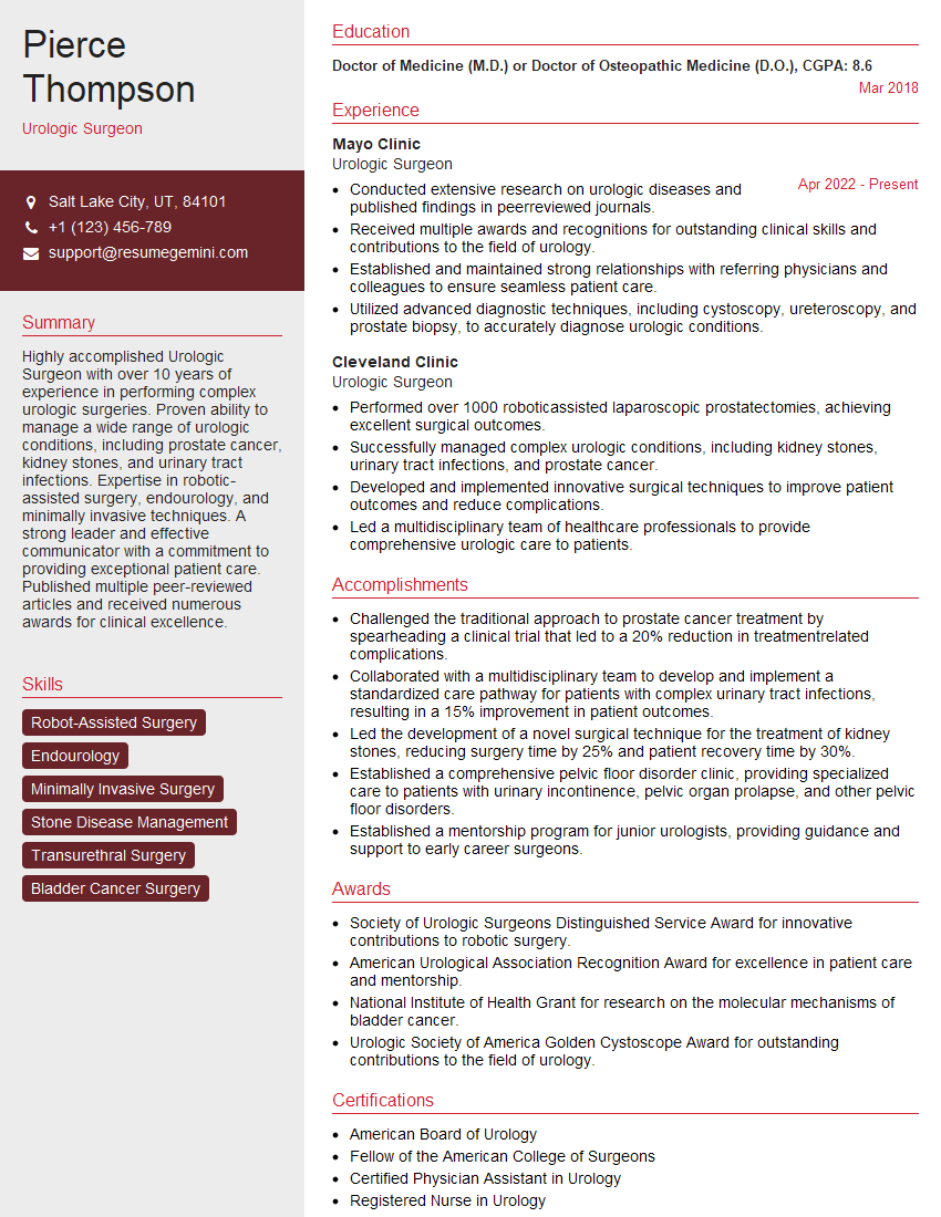
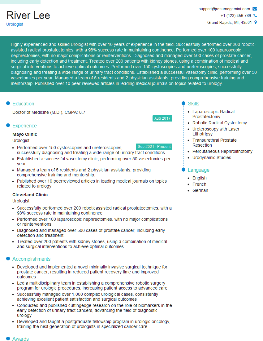
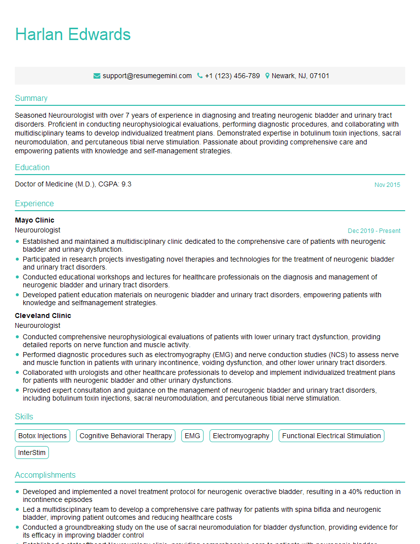
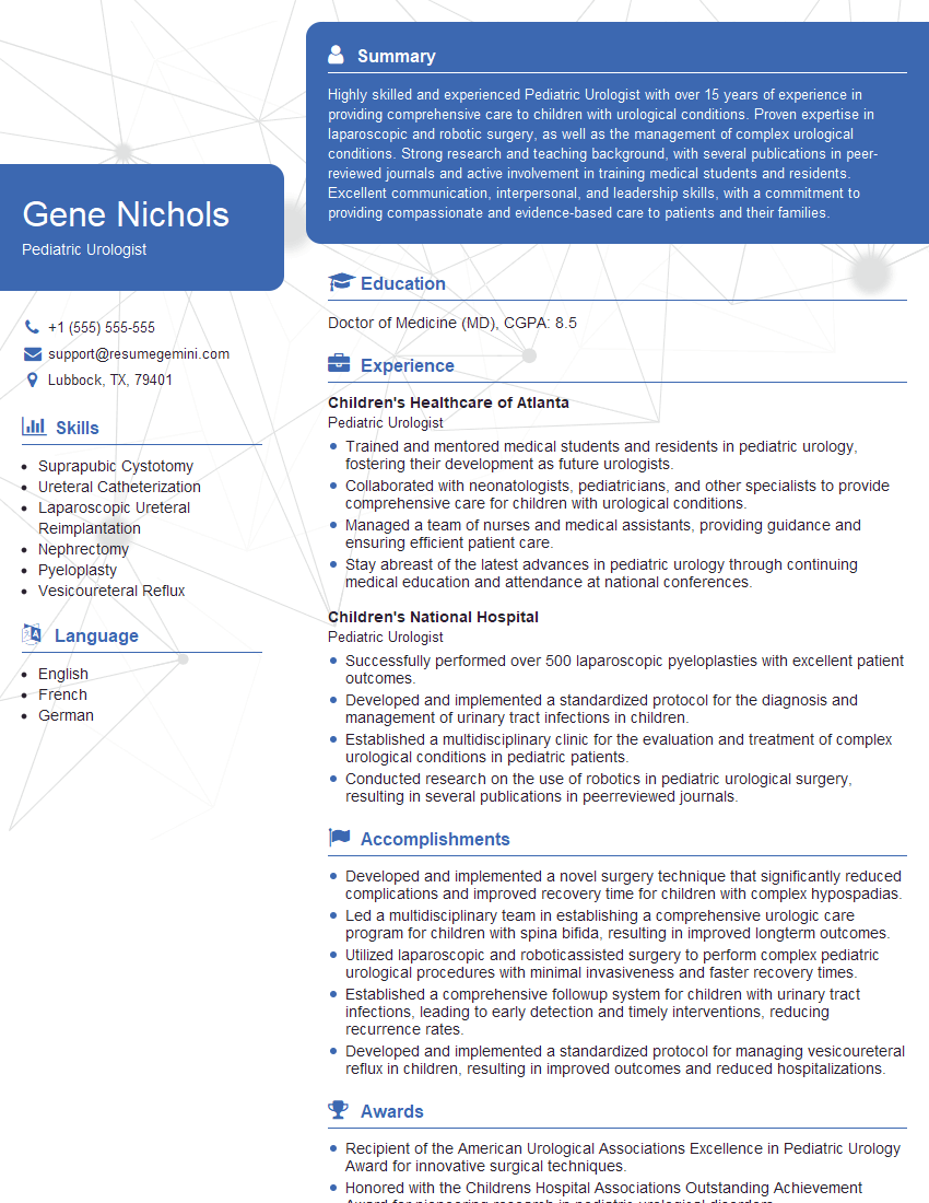
Mastering these key areas of urologic surgery is crucial for career advancement and securing your dream position. A strong foundation in both theoretical knowledge and practical application will significantly enhance your interview performance. To maximize your job prospects, create an Applicant Tracking System (ATS)-friendly resume that highlights your skills and experience effectively. ResumeGemini is a trusted resource to help you build a professional and impactful resume, ensuring your qualifications are clearly presented to potential employers. Examples of resumes tailored to Urologic Surgery are available to guide you.
Explore more articles
Users Rating of Our Blogs
Share Your Experience
We value your feedback! Please rate our content and share your thoughts (optional).
What Readers Say About Our Blog
Take a look at this stunning 2-bedroom apartment perfectly situated NYC’s coveted Hudson Yards!
https://bit.ly/Lovely2BedsApartmentHudsonYards
Live Rent Free!
https://bit.ly/LiveRentFREE
Interesting Article, I liked the depth of knowledge you’ve shared.
Helpful, thanks for sharing.
Hi, I represent a social media marketing agency and liked your blog
Hi, I represent an SEO company that specialises in getting you AI citations and higher rankings on Google. I’d like to offer you a 100% free SEO audit for your website. Would you be interested?


